Fig. 1.

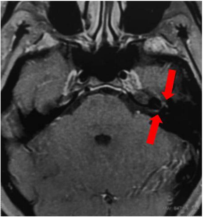
Axial brain MRI (T1/gadolinium) showing contrast enhancement in the distal intracanalicular portion in the tympanic and mastoid segments of the left facial nerve (red arrows)

Axial brain MRI (T1/gadolinium) showing contrast enhancement in the distal intracanalicular portion in the tympanic and mastoid segments of the left facial nerve (red arrows)