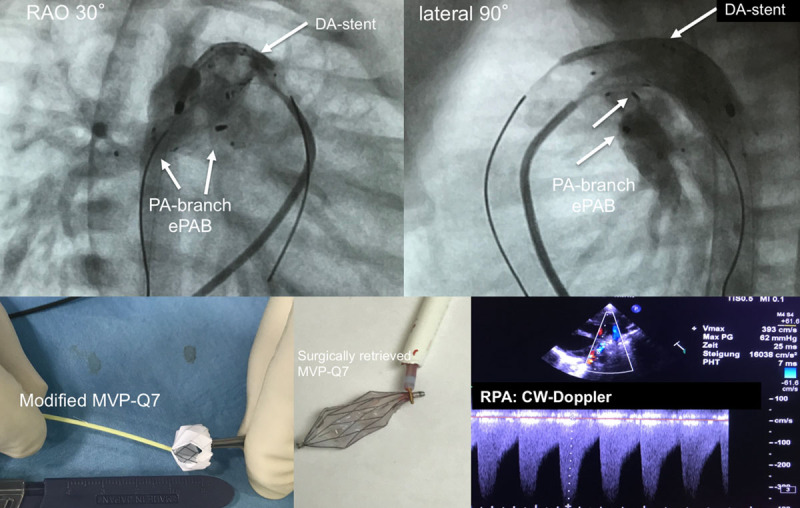
Figure.

Transcatheter stage-I. Completion angiographies are shown in right anterior oblique (RAO 30°) and lateral 90° plane after placement of modified Micro Vascular Plug within the pulmonary branches and stented duct. Furthermore, a modified MVP-7Q is depicted after removal of polytetrafluoroethylene from 2 segments, as well as a device after intraoperative removal by the snare technique. In addition, a 2D echocardiographic short-axis plane is depicted, with continuous-wave (CW) Doppler tracing representing pulmonary systolic-diastolic flow profile of an effective pulmonary flow restrictor obtained from the right pulmonary artery (RPA). DA indicates arterial duct; and PA, pulmonary artery.
