
Figure 4.
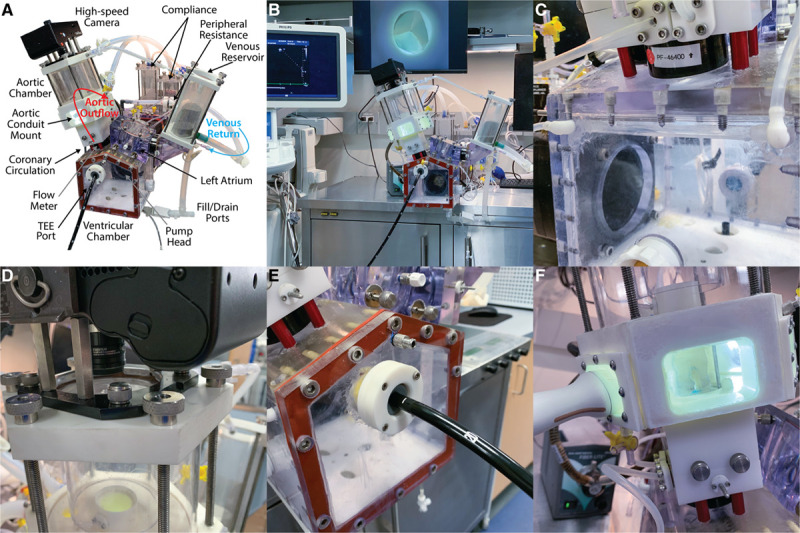
Components of the Stanford left heart simulator system. A, Left heart simulator labeled component drawing. B, Overview of the entire experimental setup, including the left heart simulator, echocardiography machine, and high-speed videometric output. Computers, flow meters, pump controllers, input/output controller, and data acquisition system are in the cabinet below and not shown. C, Close-up view of the dual differential-resistance coronary circulation with intraventricular left coronary segment replicating physiological conditions. D, High-speed camera mounted to the top of the aortic root and aortic assembly. E, View of transesophageal echocardiography (TEE) port within the left ventricular chamber for analysis. F, Transthoracic echocardiography port on the side of the aortic root enclosure for obtaining accurate in-plane images of the cusps.