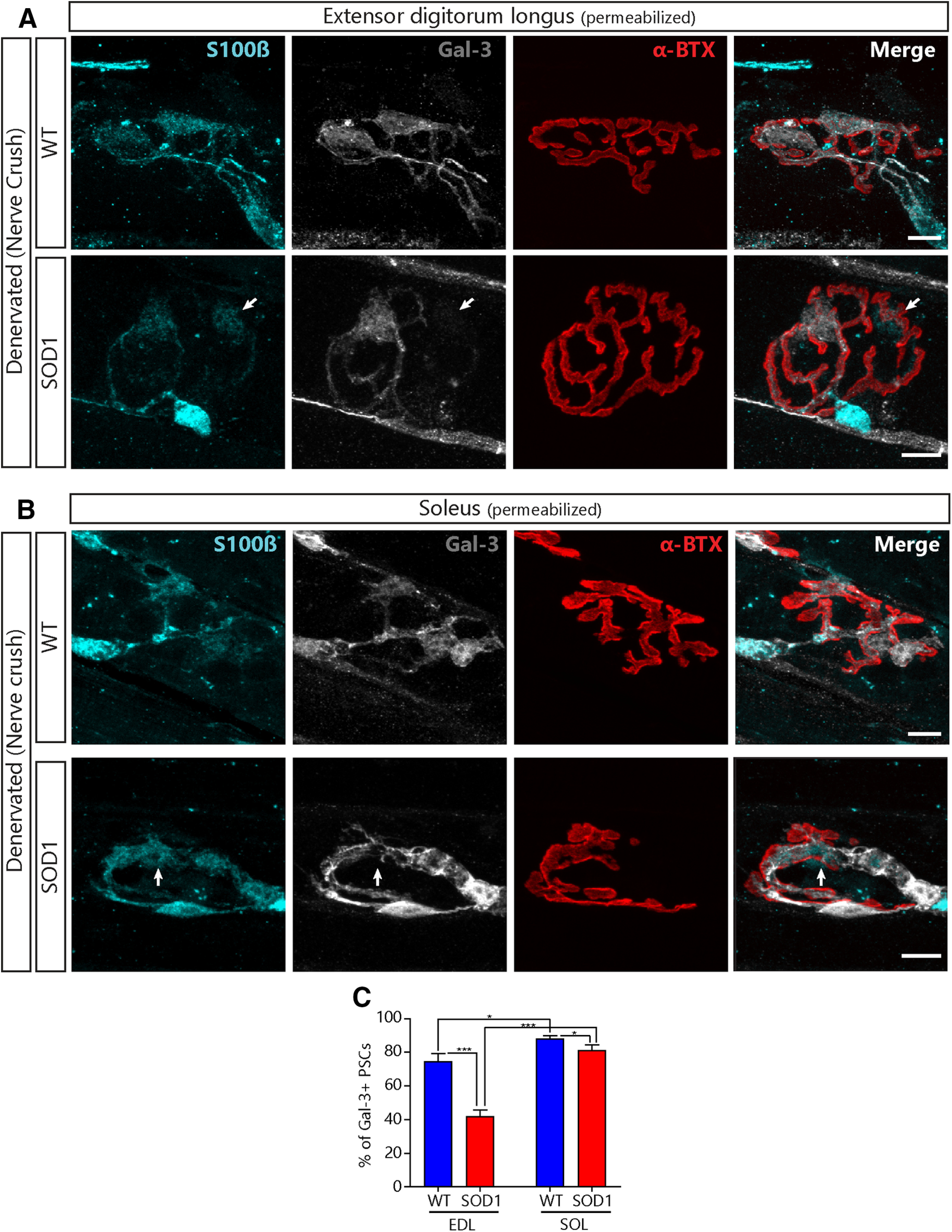
Figure 7.

Lower expression of Gal-3 in PSCs of presymptomatic SOD1G37R mice following denervation. Representative confocal images of the immunolabeling of glial cells (blue; S100β), Gal-3 (white), and postsynaptic nAChRs (red, α-BTX) from the EDL (A) or the SOL (B) of WT (top) and SOD1G37R mice (bottom) 2 after a sciatic nerve crush. Note the absence of Gal-3 expression in some of the PSCs from SOD1 mice (arrows). C, Quantification of the percentage of PSCs expressing Gal-3 2 d following a sciatic nerve crush as a function of the genotype and the muscle type. Scale bar = 10 μm; *p < 0.05, ***p < 0.001.
