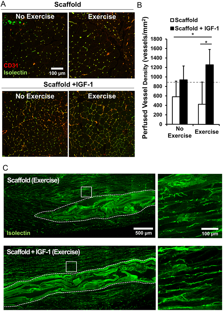
Figure 4.

Vessel regeneration and perfusion. (A) Immunofluorescence staining of co-stained CD31+/Isolectin+ anastomotic vessels in the TA muscle (B) Quantified density of perfused vessels within a 500 μm area from the scaffold region. Dotted line represents control non-regenerating tissue values (C) Characterize of the vascular network along the myofiber bundles using immunofluorescence staining of Isolectin perfused vessels in longitudinal muscle fibers adjacent to scaffold implant. (n=4 for each group except n=6 for the scaffold+IGF-1 with exercise). * denotes a statistically significant relationship (p<0.05).
