PLOS ONE logoLink to PLOS ONE
. 2020 Oct 2;15(10):e0240109. doi: 10.1371/journal.pone.0240109

Comparison of lamina cribrosa properties and the peripapillary vessel density between branch retinal vein occlusion and normal-tension glaucoma

Je Moon Woo 1, Jae Bong Cha 1, Chang Kyu Lee 1,2,*
Editor: Sanjoy Bhattacharya3
PMCID: PMC7531791  PMID: 33007029

Abstract

Purpose

To compare the properties of the lamina cribrosa (LC) and the peripapillary vessel density between branch retinal vein occlusion (BRVO) and normal-tension glaucoma (NTG), using swept-source optical coherence tomography and optical coherence tomography angiography.

Methods

This retrospective study included 21 eyes of 21 patients with BRVO and 43 eyes of 43 patients with NTG who were treated from June 2016 to September 2017. The anterior LC depth (ALCD) and LC thickness (LCT) at the mid-superior, central, and mid-inferior levels; the mean difference in ALCD; and the peripapillary vessel density in the superficial and deep capillary plexuses and the choriocapillaris were compared between groups.

Results

ALCD at the mid-superior, central, and mid-inferior levels was significantly greater in the NTG group (P < 0.05), while LCT was comparable between the groups. The mean difference in ALCD was significantly greater in the BRVO group (P = 0.03). The peripapillary vessel density in the superotemporal segment of the superficial capillary plexus was significantly lower in the BRVO group, while the density in all segments of the choriocapillaris was significantly lower in the NTG group (P < 0.05 for all).

Conclusions

Our findings demonstrate that BRVO and NTG have different LC structures and peripapillary vessel densities.

Introduction

Retinal vein occlusion is a common cause of vision loss after diabetic retinopathy and age-related macular degeneration [1, 2]. It has two forms: central retinal vein occlusion and branch retinal vein occlusion (BRVO). BRVO is associated with a higher prevalence of arterial hypertension, peripheral vascular disease, venous disease, and peptic ulcer [3]. Some patients with BRVO have clinical signs, such as optic disc hemorrhage, an increased cup-to-disc ratio, and visual field defects, that are commonly found in glaucoma patients [4]. This suggests that BRVO and glaucoma, particularly normal-tension glaucoma (NTG), may have a similar pathological mechanism such as blood flow insufficiency, although NTG is more often associated with status of hypotension, headache and generalized vasoconstriction of blood vessels especially on the extremities [58]. Moreover, some cases of chronic BRVO are difficult to distinguish from those of NTG because of the similar topographical and morphological characteristics [8, 9]. However, the clinical prognoses of BRVO and NTG are different. While BRVO remains stable, unless there is a recurrence of retinal embolism, NTG is a progressive optic neuropathy.

Accordingly, comparisons of the ocular regions primarily affected in these two conditions, i.e., the lamina cribrosa (LC) and peripapillary vessels [10, 11], may be key to differentiating these conditions and may provide insight into the pathogenesis of NTG. To our knowledge, no study to date has performed such comparisons because of the low penetration and detection abilities of conventional devices and the prohibitive invasiveness of existing equipment required for the evaluation of these sites. However, the availability of swept-source (SS) optical coherence tomography (OCT), one of the latest noninvasive, high-speed imaging techniques, using longer wavelengths, has increased in recent years. This technique allows better imaging of posterior eye structures, such as the LC, than does enhanced-depth imaging OCT [12]. OCT angiography (OCTA) is another new imaging technique that allows visualization of all layers of the retinal and choroidal microvasculature, without requiring dye injection [13].

The aim of the present study was to evaluate the differences in the properties of the LC and the peripapillary vessel density between BRVO and NTG, using SS-OCT and OCTA, in order to provide a deeper understanding of the pathogenesis of NTG.

Materials and methods

Subjects

This retrospective cohort study adhered to the tenets of the Declaration of Helsinki and was approved by the Ulsan University Hospital Institutional Review Board (UUH 2017-09-037-001). All subjects were enrolled between June 2017 and September 2018. Our IRB committee waived the requirement for informed consent, due to the retrospective nature of the study and the full anonymity of the data before we accessed them.

Patients with NTG were recruited from the Department of Ophthalmology at Ulsan University Hospital, South Korea, using the following inclusion criteria: availability of an average of three intraocular pressure measurements obtained using Goldmann applanation tonometry at different time points, < 21 mmHg; cup-to-disc ratio for each eye of ≥ 0.5 or a difference between the two eyes of > 0.2; a defect in the retinal nerve fiber layer (consistent with a glaucomatous change in the optic nerve) observed on a fundus photograph taken under red-free light or on an optical coherence tomography image; evidence of glaucomatous visual field loss observed using a Humphrey Field Analyzer (with the Swedish Interactive Threshold Algorithm-Standard 30–2 or 24–2 program); and confirmation of open-angle glaucoma by gonioscopic examination or the Van Herick method.

NTG eyes were selected by the following criteria: 1) eyes of patients with unilateral NTG; 2) in case of bilateral NTG, the eye with higher resolution OCT image; and 3) in case of similar resolutions of the OCT images in patients with bilateral NTG, the right eye was selected to minimize selection bias.

Age-matched patients with BRVO were enrolled from the Retinal Clinic at Ulsan University Hospital, South Korea. The presence of BRVO was determined based on ophthalmoscopic slit-lamp fundus examinations, color fundus photography and OCT tomography [14]. BRVO was identified when a localized area of the retina exhibited scattered superficial and deep retinal hemorrhage, venous dilation, intraretinal microvascular abnormalities, and occluded and sheathed retinal venules. Additional inclusion criteria were as follows: intraocular pressure < 21 mmHg; minimum 6-month follow-up for stabilizing the retinal nerve fiber layer thickness [15]; and no laser therapy during the follow-up period, in order to exclude the effects of this therapy on the retinal nerve fiber layer and peripapillary vessels.

The exclusion criteria were as follows: primary open-angle glaucoma diagnosed before or during the follow-up period; best-corrected visual acuity < 0.1 on a decimal scale or > 1.0 on the logarithm of the minimum angle of resolution scale; presence of uveitis or other retinal diseases; laser therapy or intraocular surgeries, such as cataract surgery or vitrectomy, during the follow-up period; myopia of ≥ −6 D; and a history of cerebrovascular disease, ocular trauma, or any other condition that could affect the retinal nerve fiber layer and LC.

SS-OCT imaging of the optic nerve head

The optic nerve in each patient was imaged using a commercially available SS-OCT device (DRI OCT Triton, Topcon, Japan) with a wavelength of 1050 nm and a swept source with a tuning range of 100 nm for optic nerve head scanning. The device produces scans with a axial resolution of 8 μm and a transverse resolution of 20 μm. This instrument provides high-quality images with constant signal strength throughout the posterior pole, even in eyes with cataracts that affect image quality [16]. Moreover, the instrument performs up to 100,000 A-scans per second and has an invisible scanning line because of the long wavelength; this can reduce eye movement and ensure more successful scans. We obtained images using a 6 × 6-mm area with a depth of 2.6 mm centered on the optic nerve head. Each dataset comprised 256 cross-sectional B-scan images of 512 × 256 pixels. For inclusion in the analyses, all images were required to have an image quality score of ≥ 35 according to the manufacturer’s recommendations.

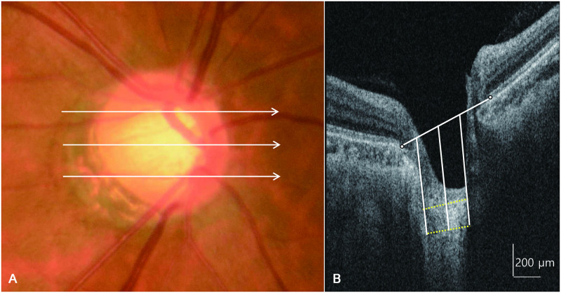
For identification of the anterior and posterior borders of the LC, horizontal SS OCT B-scans were assessed using Adobe Photoshop CS2 (version 9.0; Adobe Systems, Inc., San Jose, California, USA). The LC thickness (LCT) was defined as the distance between the anterior and posterior borders of the highly reflective region seen beneath the optic nerve head on a cross-sectional B-scan image (Fig 1).

Fig 1. A fundus photograph and horizontal cross-sectional B-scan images of the optic nerve head obtained using swept-source optical coherence tomography showing measurement of lamina cribrosa thickness.

Fig 1

The lamina cribrosa thickness is measured at the mid-superior, central, and mid-inferior levels of the optic nerve head (A). At each level, the thickness is measured at three points (center, 100 μm nasally, and 100 μm temporally), along a line perpendicular to a reference line connecting the two ends of Bruch’s membrane (B).

LCT was determined using a method similar to that of Kwun et al. [17], in which measurements were obtained from three lines, i.e., the mid-superior, central, and mid-inferior lines of the optic disc, using the manual caliper tool in DRI OCT Viewer 9.01 software. The mid-superior and mid-inferior lines were identified as the horizontal line located at the halfway point on the vertical line that connects the optic disc center to the margin. At each line, LCT was measured at three locations along a perpendicular line from the reference line connecting the two ends of the Bruch membrane, i.e., at the center of the reference line, and 100-μm temporally and 100-μm nasally from this line. The center of the reference line was used for comparison between the two diseases, and the nasal and temporal lines were used as supporting values for the central reference line. Anterior LC depth (ALCD) was defined as the distance between the reference line connecting the end of the Bruch membrane and the anterior border of the LC. We also determined differences in LC evenness between BRVO and NTG and calculated the mean difference of ALCD from the mid-superior to mid-inferior portion of the central LC. A lower value indicated a more even, or flat, LC.

OCTA of the peripapillary area

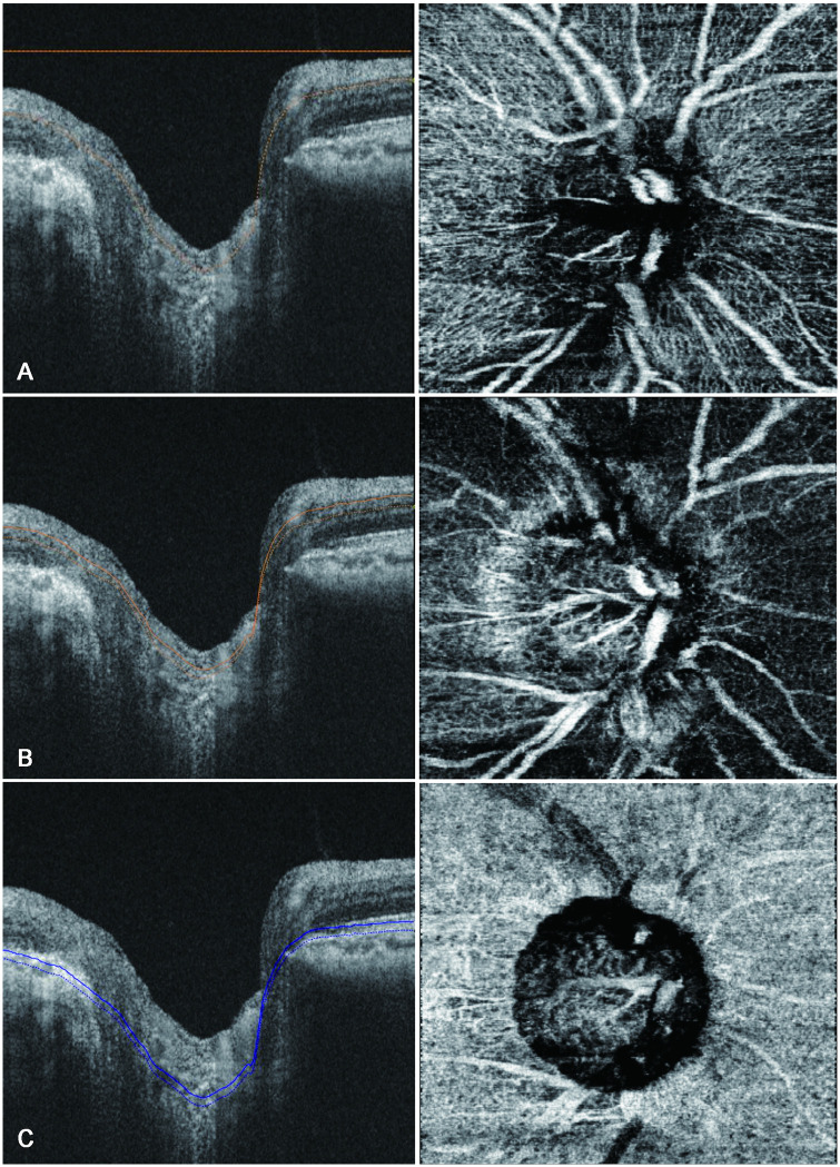
SS-OCTA was performed using the DRI-OCT triton (Topcon) to assess the structure of the peripapillary capillary plexus. Images were obtained using a 3 × 3-mm scan area centered on the optic disc. We analyzed the peripapillary capillary density in the superficial capillary plexus, which comprised the inner capillary layer of the retina; the deep capillary plexus, which comprised the outer capillary layer; as well as the choriocapillaris. The retinal vascular plexus described by Weinhaus et al. [18] was simplistically considered as two main layers, as described by Snodderly et al. [19] The inner capillary layer or the superficial capillary plexus started from the internal limiting membrane region and was of sufficient thickness to include the larger arteries, arterioles, capillaries, venules, and veins in the ganglion cell layer. It ended at the inner plexiform, which is the most superficial hyporeflective band. The outer capillary layer or the deep capillary plexus was identified by segmenting en-face images such that the inner boundary coincided with the inner nuclear layer and the outer boundary coincided with the midpoint of the outer plexiform layer [20, 21]. The choriocapillaris was identified by a 10-μm thick slab offset of 21 μm below the instrument generating the retinal pigment epithelial band (Fig 2) [22]. We used latest built-in software (IMAGEnet6) to generate OCT-angiograms which can provide improved detection sensitivity of low blood flow and reduced motion artifacts without compromising axial resolution [23]. Included OCTA images were subsequently exported in PNG format and imported into an i-solution image analysis program (iMT, i-Solution Inc).

Fig 2. B-scan images of the optic nerve (left) and peripapillary vessels (right) obtained using swept-source optical coherence angiography showing measurement of peripapillary vessel density.

Fig 2

The peripapillary vessel density is measured in the superficial capillary plexus, which comprises the inner capillary layer of the retina (A); the deep capillary plexus, which makes up the outer capillary layer (B); and the choriocapillaris (C).

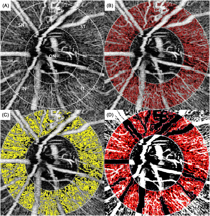
Subsequently, each of the three layers was segmented into superotemporal, inferotemporal, superonasal, and inferonasal regions. Image processing and analysis consisted of 3 stages [24]. The first stage was extraction of the region of interest (ROI) around the optic nerve. The optic nerve was identified as a circle, and the outer margin was placed at 750 μm from the margin of this circle. In cases of myopia with a zone of peripapillary chorioretinal atrophy, the circle included the zone of atrophy, to minimize the influence on the peripapillary vessel density [25, 26]. The donut-shaped portion between the outer margin and the optic nerve margin was divided into four segments using Adobe Photoshop CS2 (Adobe Systems, Inc.), and finally, the donut-shaped ROI was extracted (Fig 3A and 3B). The second stage was detection and deletion of thick vessels from the ROI. We checked large vessels such as the inferior and superior nasal and the inferior and superior temporal retinal veins and arteries, and we excluded these vessels one-by-one using sub mode (deletion mode) with a built-in program in i-solution (Fig 3C and 3D). The third stage was estimation of the peripapillary vessel density, which is the ideal measure of capillaries per unit area. The desired image is the binary image, in which white pixels represent capillaries (without large vessels) and black pixels represent non-vessels. The peripapillary vessel density was calculated using the following formula: peripapillary vessel density (%) = (Nw / A) × 100, where Nw represents the number of white pixels and A represents the area of the selected image sector. As both the numerator and the denominator are pixel counts, the peripapillary vessel density is reported between 0% and 100% [2730].

Fig 3. Segmental valuation of the peripapillary vessel density.

Fig 3

The optic nerve was indicated as a circle, and the outer margin was set at 750 μm from this circle margin. The donut-shaped portion between the outer margin and the optic nerve margin was divided into four segments, namely the superotemporal, inferotemporal, superonasal, and inferonasal segments (A); each segment was extracted as a region of interest (ROI) (B); large vessels were detected and deleted (C); and finally, the binary ROI of the peripapillary vessels, with large vessels excluded, was retained (D). ON, optic nerve; ST, superotemporal; IT, inferotemporal; SN, superonasal; IN, inferonasal.

Data analysis

Within-visit repeatability of the peripapillary vessel density measurements was calculated using two sets of images that were sequentially obtained in a single visit. The interobserver reproducibility was calculated from measurements recorded for 10 randomly selected eyes by two independent observers (JBC and JMW) who were masked to the patients’ information. Variability was assessed using the intraclass correlation coefficient (ICC) with a two-way random effects model.

The two groups were matched for age and the mean deviation prior to performing intergroup comparisons, for which independent t-tests were used. In analyzing peripapillary vessel density, we used one-way multivariate analysis of variance. All statistical analyses were performed using Statistical Package for the Social Sciences (SPSS) for Windows (version 21.0, SPSS, Inc., Chicago, IL, USA). A P-value of < 0.05 was considered statistically significant.

Result

A total of 71 patients were initially included in this study. Of these, three were excluded from the BRVO group, since one patient showed the possibility of primary open-angle glaucoma concomitant with BRVO and the other two had poor-quality OCTA images. Four patients with poor-quality OCTA images were also excluded from the NTG group. Eventually, 21 eyes of 21 patients with BRVO and 43 eyes of 43 patients with NTG were analyzed. The age range of patients overall was 48–79 years (mean age: 64.81 ± 8.81 years). There were no significant differences in age, sex, and laterality between the two groups (P = 0.79, P = 0.42, and P = 0.30, respectively). Moreover, the two groups showed no significant differences in terms of a history of hypertension or diabetes mellitus and Humphrey visual field indices, such as the mean deviation and pattern standard deviation (Table 1).

Table 1. Baseline characteristics of BRVO and NTG patients.

BRVO group (n = 21) NTG group (n = 43) P value
Age, year (mean ± SD) 58.19 ± 10.75 57.39 ± 13.23 0.79
Sex, men/women (% (n)) 47 (10)/53 (11) 58.1 (25)/41.9 (18) 0.42
Laterality (OD/OS) 6/15 18/25 0.3
S.E. (diopter) -1.25± 0.85 -2.45±1.12 0.3
HTN (%) 42.8 25.5 0.16
DM (%) 9.5 6.9 0.72
HVF
 MD (dB) -8.93 ± 5.17 -8.61 ± 7.9 0.84
 PSD (dB) 8.94 ± 2.68 7.60 ± 4.04 0.17

*P < 0.05.

BRVO: branch retinal vein occlusion, NTG, normal tension glaucoma; S.E.: Spherical equivalent, HTN: hypertension, DM: diabetic mellitus, HVF: Humphrey visual field, MD: mean deviation, PSD: pattern standard deviation, dB: decibel.

Comparison of LC properties

The ALCD at the mid-superior, central, and mid-inferior levels was significantly greater in the NTG group than in the BRVO group (P = 0.02, 0.01, and 0.01, respectively; Table 2).

Table 2. Comparing anterior lamina depth between BRVO and NTG patients.

BRVO group (n = 21) NTG group (n = 43) P value
Mid-superior ALDn, μm 329.4 ± 122.2 378.3 ± 141.6 0.23
ALDc, μm 366.7 ± 119.9 450.9 ± 138.0 0.02*
ALDt, μm 223.5 ± 74.9 320.6 ± 123.1 0.02*
Central ALDn, μm 301.4 ± 106.2 345.7 ± 129.4 0.22
ALDc, μm 322.2 ± 105.7 432.5 ± 134.0 0.01*
ALDt, μm 271.6 ± 107.6 309.5 ± 127.1 0.29
Mid-inferior ALDn, μm 273.9 ± 93.8 301.2 ± 116.1 0.35
ALDc, μm 292.9 ± 115.1 406.0 ± 123.0 0.01*
ALDt, μm 264.0 ± 100.3 298.5 ± 109.0 0.22

*P < 0.05.

BRVO: branch retinal vein occlusion, NTG: normal tension glaucoma, ALDn: anterior lamina depth of nasal side, ALDc: anterior lamina depth of central side, ALDt: anterior lamina depth length of temporal side.

LCT at all levels was smaller in the NTG group than in the BRVO group, although the differences were not significant at any level (Table 3).

Table 3. Comparing lamina cribrosa thickness between BRVO and NTG patients.

BRVO group (n = 21) NTG group (n = 43) P value
Mid-superior LCn, μm 175.8 ± 75.6 169.2 ± 47.9 0.67
LCc, μm 177.9 ± 78.8 165.5 ± 56.7 0.47
LCt, μm 182.0 ± 56.28 157.9 ± 52.0 0.10
Central LCn, μm 183.4 ± 63.3 173.2 ± 53.2 0.52
LCc, μm 176.9 ± 57.4 152.7 ± 52.9 0.11
LCt, μm 186.3 ± 81.8 170.7 ± 65.7 0.45
Mid-inferior LCn, μm 176.8 ± 38.2 175.6 ± 53.1 0.91
LCc, μm 178.5 ± 70.5 164.3 ± 65.3 0.44
LCt, μm 172.9 ± 48.0 165.1 ± 62.5 0.58

*P < 0.05.

BRVO: branch retinal vein occlusion, NTG: normal tension glaucoma, LCn: lamina cribrosa thickness of nasal side, LCc: lamina cribrosa thickness of central side, LCt: lamina cribrosa thickness of temporal side.

In the BRVO group, the superotemporal segment was the site of retinal vein occlusion in 16 patients (77%), the inferotemporal segment was affected in 4 (19%) and both the supero- and inferotemporal segments were affected in one patient (4%).

The mean difference in ALCD from the mid-superior to the mid-inferior levels of the central LC was significantly greater in the BRVO group than in the NTG group (74.8 ± 39.1 μm and 41.6 ± 57.1 μm, respectively; P = 0.03; Table 4).

Table 4. Mean difference of anterior lamina depth from mid-superior to mid-inferior portion of central lamina cribrosa.

Mean difference BRVO group (n = 21) NTG group (n = 43) P value
ALCD, μm 74.8 ± 52.8 41.7 ± 46.7 0.02*

*P < 0.05.

BRVO: branch retinal vein occlusion, NTG: normal tension glaucoma.

Comparisons of the peripapillary capillary density

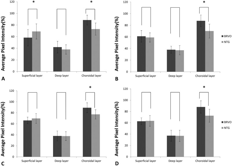
The peripapillary capillary density in the superotemporal segment of the superficial capillary plexus was significantly lower in the BRVO group than in the NTG group (58.25 ± 14.24 and 68.90 ± 13.15, respectively; P = 0.005; Fig 4A). For all segments in the choriocapillaris, the vessel density was significantly lower in the NTG group than in the BRVO group (P < 0.05 for all; Fig 4). The vessel density in the inferotemporal segment of the superficial capillary plexus was lower in the NTG group than in the BRVO group, although the difference was not statistically significant (P = 0.401; Fig 4B). The NTG group generally tended to exhibit a lower vessel density in all segments in the deep layer than did the BRVO group; however, these differences were not statistically significant (Fig 4).

Fig 4. Swept-source optical coherence angiography findings for the peripapillary vessel density in different layers in eyes with branch retinal vein occlusion (BRVO) and those with normal-tension glaucoma (NTG) (A, Superiotemporal; B, Inferotemporal; C, Superonasal; D Inferonasal area).

Fig 4

The vessel density in the superotemporal segment of the superficial capillary plexus is significantly lower in BRVO than in NTG (P = 0.008) (A). The vessel density in all segments in the choriocapillaris is significantly lower in NTG than in BRVO (P < 0.05) (A–D) (Error bar = standard deviation).

Repeatability and reproducibility

For peripapillary vessel density measurements, ICCs for within-visit repeatability and interobserver reproducibility with the i-solution® program were 0.956 and 0.975 (excellent), respectively.

Discussion

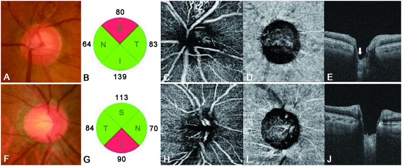
In this study, we hypothesized that LC properties and peripapillary vessel density would differ between BRVO and NTG patients, and that this could help differentiate between the pathogeneses of the two conditions. Accordingly, we used SS-OCT and SS-OCTA to investigate this hypothesis; we indeed found that such differences exist (Fig 5).

Fig 5.

Fig 5

Lamina cribrosa properties and the peripapillary vessel density for representative cases of branch retinal vein occlusion (BRVO) and normal-tension glaucoma (NTG) (A–E). The left eye of a patient with BRVO, showing a mean deviation (Humphrey visual field [HVF]) of −5.21 dB (F–J). The right eye of a patient with NTG, showing a mean deviation (HVF) of −5.41 dB. The eye with BRVO shows superior disc notching (A) and a superior retinal nerve fiber layer (RNFL) defect (B). The peripapillary vessel density in the superotemporal segment of the superficial capillary plexus has decreased (C), with more superior disc cupping seen on a vertical B-scan (white arrow, E). The vessel density in the choriocapillaris is lower in the eye with NTG (I) than in the eye with BRVO. Moreover, the eye with NTG shows greater disc cupping and a more uniform LC (J) than does the eye with BRVO.

ALCD was generally greater in the NTG than in the BRVO group, while LCT was insignificantly smaller in the former. Moreover, the LC was more irregular in BRVO, with its superior portion being the most common site of retinal vein occlusion. The optic nerve head is considered a biomechanically weak area because it coincides with discontinuity in the corneoscleral shell [31]. The LC is a mesh-like structure that is composed of a multilayered network of collagen fibers and occupies a hole in the sclera. It allows the nerve fibers of the optic nerve and retinal vessels to pass through the sclera and to exit the eye. Therefore, LCT can decrease in cases with ganglion cell degeneration and axonal damage with consequent inhibition of axonal transport [32, 33]. The mechanism involved is similar to that by which a column in a concrete building weakens upon removal of the reinforcing bars. The most typical disease associated with ganglion cell degeneration and axonal damage is glaucoma, particularly NTG. Several studies have reported that the LC and pre-LC are thinner in eyes with NTG than in healthy eyes [3436]. Kim et al. [37] found that the anterior laminar insertion depth was greater than normal in eyes with NTG. The LC and pre-LC were also found to be thinner in eyes with BRVO, due to axonal damage and shallow optic disc excavation, than in healthy eyes [38]. However, in BRVO, which derives its name from the presence of occlusion in only a particular segment of the retinal vein, there is a tendency for the retinal nerve fiber layer to exhibit thinning only in the affected portion, while the unaffected portion of the same optic nerve does not show thinning [15]. A possible mechanism for this type of defect is that the retinal ischemic lesion may lead to focal axonal dysfunction and cause descending retinal ganglion cell degeneration and atrophy [39]. Moreover, BRVO predominantly occurs in the superotemporal segment [40], whereas the damage associated with glaucoma first involves the axonal bundles, with somewhat greater involvement of the inferior and superior poles of the optic disc and more diffuse and generalized damage than that observed in BRVO [41]. Consequently, we observed less LC thinning and disc cupping in our BRVO group than in our NTG group. Moreover, in eyes with BRVO, ALCD in the superior portion of the LC, which is the predominant site for retinal vein occlusion [40], is greater than that in the inferior portion. On the other hand, the LC shows overall uniformity in eyes with NTG (Fig 5E and 5J).

OCTA has inherent advantages due to its capacity for optical dissection and visualization of the blood flow in various layers of the retina [20]. Using OCTA, we have demonstrated a difference in the peripapillary vessel density between NTG and BRVO patients and showed that, in the BRVO group, the vessel density in the superotemporal segment (where vessel occlusion was more frequent [40]) of the superficial capillary plexus was significantly lower than that in the NTG group (Fig 4A). This could be associated with a decrease in the peripapillary retinal microvasculature at the site of the retinal nerve fiber layer defect [13, 42]. The vessel density in the inferotemporal segment (which is more frequently affected in NTG patients) of the superficial capillary plexus was lower in NTG than in BRVO patients, although the difference was not significant. In the choriocapillaris, the peripapillary vessel density in all segments was significantly lower in NTG than in BRVO patients (Figs 4, 5D and 5I), probably because of the unique blood supply in the optic nerve head. The surface nerve fiber layer is primarily supplied by retinal arterioles, and the ganglion cell layer is primarily supplied by the inner vascular plexus of the retina [43]. The choroidal layer of the peripapillary vessels supplies the prelaminar region; some of these vessels form the arterial circle of Zinn–Haller and supplies the LC region [44, 45]. Therefore, in BRVO, secondary damage to ganglion cells and the retinal nerve fiber layer may occur around the primary injured vessel, with a consequent decrease in peripapillary capillaries. On the other hand, the LC is thought to be the primary affected site in NTG. Accordingly, we speculate that, not only the density of the peripapillary capillaries (which supply consequently damaged ganglion cells and retinal nerve fibers), but also that associated with the LC, is decreased in NTG. The decrease in superficial peripapillary capillaries in BRVO may be solely due to damage to the ganglion cells and retinal nerve fiber layer. However, the decrease in the choriocapillaris vessel density in NTG may not be associated with this type of damage; rather, it may be associated with the etiology of NTG. Both BRVO and NTG present similar clinical pictures, with ganglion cell and nerve fiber layer thinning and disc atrophy [46]; yet the blood supply to the LC is significantly decreased only in NTG. This insufficient blood supply and vascular dysregulation may induce damage to the retinal nerve fibers, which pass through the LC, with consequent damage to secondary ganglion cells. Accordingly, we assume that NTG is directly caused by a decrease in the choriocapillaris vessel density, which is consistent with several studies that have indirectly implicated a low perfusion pressure and vascular dysregulation as important events in the pathogenesis of NTG [4749].

This study hads some limitations. First, blood flow quantification was not possible with our OCTA device, and we were unable to determine the precise blood flow in the peripapillary vessels. Second, our OCT device does not provide software for automatic calculations of vessel density. However, we used i-solution® software, which has been successfully used for similar calculations in other fields; moreover, the interobserver reproducibility obtained using this program was excellent. Third, the most commonly affected sites differed between the two conditions. Therefore, to increase understanding of differences in the pathogenesis of BRVO and NTG, the differences between these two conditions in the same pathological location, such as the inferotemporal or superotemporal segment, should be analyzed further. Finally, although choroidal vessels comprise the choriocapillaris, Sattler’s layer, and Haller’s layer, SS-OCTA cannot clearly depict whole choroidal vessels, unlike laser speckle flowmetry [50]. We therefore considered the choriocapillaris as representing choroidal blood flow as a whole.

Another limitation may be the different number of patients between the two groups.

In conclusion, we demonstrated that BRVO and NTG have different LC properties and peripapillary vessel densities. LC cupping is more pronounced in NTG than in BRVO, and focal cupping is more common in BRVO than in NTG, due to the focal damage to retinal nerve fibers passing through the LC in BRVO. BRVO commonly exhibits decreased capillaries in the superotemporal segment of the superficial capillary plexus, whereas NTG commonly exhibits decreased vessels in the choriocapillaris. Finally, deformation and thinning of the LC can be due to damaged and lost axonal tissue and inadequate blood supply to the LC, caused by glaucomatous damage, particularly in NTG. Further study of diseases that are clinically similar to NTG could enhance our understanding of the pathogenesis of glaucoma, particularly NTG.

Data Availability

Data are available from ulsan university hospital ethics committee (contact via 0716782@uuh.ulsan.kr/Ms. Jeong Sook Kim; secretary of ulsan university hospital institutional review board).

Funding Statement

This research was supported by Basic Science Research Program through the National Research Foundation of Korea(NRF) funded by the Ministry of Science and ICT(NRF-2017R1C1B5018279).

References

  • 1.Rogers S, McIntosh RL, Cheung N, Lim L, Wang JJ, Mitchell P, et al. The prevalence of retinal vein occlusion: pooled data from population studies from the United States, Europe, Asia, and Australia. Ophthalmology. 2010;117(2):313–9 e1. Epub 2009/12/22. 10.1016/j.ophtha.2009.07.017 [DOI] [PMC free article] [PubMed] [Google Scholar]
  • 2.Mitchell P, Liew G, Gopinath B, Wong TY. Age-related macular degeneration. Lancet. 2018;392(10153):1147–59. Epub 2018/10/12. 10.1016/S0140-6736(18)31550-2 . [DOI] [PubMed] [Google Scholar]
  • 3.Ho M, Liu DT, Lam DS, Jonas JB. Retinal vein occlusions, from basics to the latest treatment. Retina. 2016;36(3):432–48. Epub 2015/12/31. 10.1097/IAE.0000000000000843 . [DOI] [PubMed] [Google Scholar]
  • 4.Shaarawy TM, Sherwood MB, Hitchings RA, Crowston JG. Glaucoma 2nd ed Philadelphia, PA: Saunders Elsevier; 2015. [Google Scholar]
  • 5.Rath EZ, Frank RN, Shin DH, Kim C. Risk factors for retinal vein occlusions. a case-control study. Ophthalmology. 1992;99(4):509–14. Epub 1992/04/01. 10.1016/s0161-6420(92)31940-2 . [DOI] [PubMed] [Google Scholar]
  • 6.Mitchell P, Smith W, Chang A. Prevalence and associations of retinal vein occlusion in Australia. the blue mountains eye study. Arch Ophthalmol. 1996;114(10):1243–7. Epub 1996/10/01. 10.1001/archopht.1996.01100140443012 . [DOI] [PubMed] [Google Scholar]
  • 7.Hayreh SS, Zimmerman MB, Beri M, Podhajsky P. Intraocular pressure abnormalities associated with central and hemicentral retinal vein occlusion. Ophthalmology. 2004;111(1):133–41. Epub 2004/01/09. 10.1016/j.ophtha.2003.03.002 . [DOI] [PubMed] [Google Scholar]
  • 8.Beaumont PE, Kang HK. Cup-to-disc ratio, intraocular pressure, and primary open-angle glaucoma in retinal venous occlusion. Ophthalmology. 2002;109(2):282–6. Epub 2002/02/05. 10.1016/s0161-6420(01)00922-8 . [DOI] [PubMed] [Google Scholar]
  • 9.Lindblom B. Open angle glaucoma and non-central retinal vein occlusion—the chicken or the egg? Acta Ophthalmol Scand. 1998;76(3):329–33. Epub 1998/08/01. 10.1034/j.1600-0420.1998.760315.x . [DOI] [PubMed] [Google Scholar]
  • 10.Levy NS, Crapps EE. Displacement of optic nerve head in response to short-term intraocular pressure elevation in human eyes. Arch Ophthalmol. 1984;102(5):782–6. Epub 1984/05/01. 10.1001/archopht.1984.01040030630037 . [DOI] [PubMed] [Google Scholar]
  • 11.Yan DB, Coloma FM, Metheetrairut A, Trope GE, Heathcote JG, Ethier CR. Deformation of the lamina cribrosa by elevated intraocular pressure. Br J Ophthalmol. 1994;78(8):643–8. Epub 1994/08/01. 10.1136/bjo.78.8.643 [DOI] [PMC free article] [PubMed] [Google Scholar]
  • 12.Park HY, Shin HY, Park CK. Imaging the posterior segment of the eye using swept-source optical coherence tomography in myopic glaucoma eyes: comparison with enhanced-depth imaging. Am J Ophthalmol. 2014;157(3):550–7. Epub 2013/11/19. 10.1016/j.ajo.2013.11.008 . [DOI] [PubMed] [Google Scholar]
  • 13.Liu L, Jia Y, Takusagawa HL, Pechauer AD, Edmunds B, Lombardi L, et al. Optical coherence tomography angiography of the peripapillary retina in glaucoma. JAMA Ophthalmol. 2015;133(9):1045–52. Epub 2015/07/24. 10.1001/jamaophthalmol.2015.2225 [DOI] [PMC free article] [PubMed] [Google Scholar]
  • 14.Ghasemi Falavarjani K, Phasukkijwatana N, Freund KB, Cunningham ET Jr., Kalevar A, McDonald HR, et al. En Face Optical Coherence Tomography Analysis to Assess the Spectrum of Perivenular Ischemia and Paracentral Acute Middle Maculopathy in Retinal Vein Occlusion. Am J Ophthalmol. 2017;177:131–8. Epub 2017/02/27. 10.1016/j.ajo.2017.02.015 . [DOI] [PubMed] [Google Scholar]
  • 15.Kim CS, Shin KS, Lee HJ, Jo YJ, Kim JY. Sectoral retinal nerve fiber layer thinning in branch retinal vein occlusion. Retina. 2014;34(3):525–30. Epub 2013/08/21. 10.1097/IAE.0b013e3182a2e746 . [DOI] [PubMed] [Google Scholar]
  • 16.Esmaeelpour M, Povazay B, Hermann B, Hofer B, Kajic V, Kapoor K, et al. Three-dimensional 1060-nm OCT: choroidal thickness maps in normal subjects and improved posterior segment visualization in cataract patients. Invest Ophthalmol Vis Sci. 2010;51(10):5260–6. Epub 2010/05/07. 10.1167/iovs.10-5196 . [DOI] [PubMed] [Google Scholar]
  • 17.Kwun Y, Han JC, Kee C. Comparison of lamina cribrosa thickness in normal tension glaucoma patients with unilateral visual field defect. Am J Ophthalmol. 2015;159(3):512–8 e1. Epub 2014/12/17. 10.1016/j.ajo.2014.11.034 . [DOI] [PubMed] [Google Scholar]
  • 18.Weinhaus RS, Burke JM, Delori FC, Snodderly DM. Comparison of fluorescein angiography with microvascular anatomy of macaque retinas. Exp Eye Res. 1995;61(1):1–16. Epub 1995/07/01. 10.1016/s0014-4835(95)80053-0 . [DOI] [PubMed] [Google Scholar]
  • 19.Snodderly DM, Weinhaus RS, Choi JC. Neural-vascular relationships in central retina of macaque monkeys (Macaca fascicularis). J Neurosci. 1992;12(4):1169–93. Epub 1992/04/01. 10.1523/JNEUROSCI.12-04-01169.1992 . [DOI] [PMC free article] [PubMed] [Google Scholar]
  • 20.Spaide RF, Klancnik JM Jr., Cooney MJ. Retinal vascular layers imaged by fluorescein angiography and optical coherence tomography angiography. JAMA Ophthalmol. 2015;133(1):45–50. Epub 2014/10/16. 10.1001/jamaophthalmol.2014.3616 . [DOI] [PubMed] [Google Scholar]
  • 21.Campbell JP, Zhang M, Hwang TS, Bailey ST, Wilson DJ, Jia Y, et al. Detailed vascular anatomy of the human retina by projection-resolved optical coherence tomography angiography. Sci Rep. 2017;7:42201 Epub 2017/02/12. 10.1038/srep42201 significant financial interest in Optovue, Inc., a company that may have a commercial interest in the results of this research and technology. These potential conflicts of interest have been reviewed and managed by OHSU. David Huang receives patent royalties from Carl Zeiss Meditec, Inc. Other authors do not have financial interest in the subject of this article. [DOI] [PMC free article] [PubMed] [Google Scholar]
  • 22.Byon I, Alagorie AR, Ji Y, Su L, Sadda SR. Optimizing the Repeatability of Choriocapillaris Flow Deficit Measurement From Optical Coherence Tomography Angiography. Am J Ophthalmol. 2020;219:21–32. Epub 2020/05/27. 10.1016/j.ajo.2020.05.027 . [DOI] [PubMed] [Google Scholar]
  • 23.Tang FY, Ng DS, Lam A, Luk F, Wong R, Chan C, et al. Determinants of Quantitative Optical Coherence Tomography Angiography Metrics in Patients with Diabetes. Sci Rep. 2017;7(1):2575 Epub 2017/06/02. 10.1038/s41598-017-02767-0 [DOI] [PMC free article] [PubMed] [Google Scholar]
  • 24.Mansoori T, Sivaswamy J, Gamalapati JS, Agraharam SG, Balakrishna N. Measurement of Radial Peripapillary Capillary Density in the Normal Human Retina Using Optical Coherence Tomography Angiography. J Glaucoma. 2017;26(3):241–6. Epub 2016/12/03. 10.1097/IJG.0000000000000594 . [DOI] [PubMed] [Google Scholar]
  • 25.Lee EJ, Lee KM, Lee SH, Kim TW. Parapapillary choroidal microvasculature dropout in glaucoma: a comparison between optical coherence tomography angiography and indocyanine green angiography. Ophthalmology. 2017;124(8):1209–17. Epub 2017/04/24. 10.1016/j.ophtha.2017.03.039 . [DOI] [PubMed] [Google Scholar]
  • 26.Kiyota N, Kunikata H, Takahashi S, Shiga Y, Omodaka K, Nakazawa T. Factors associated with deep circulation in the peripapillary chorioretinal atrophy zone in normal-tension glaucoma with myopic disc. Acta Ophthalmol. 2018;96(3):e290–e7. Epub 2017/11/25. 10.1111/aos.13621 . [DOI] [PubMed] [Google Scholar]
  • 27.Oh SH, Park IK, Kim JM, Lee JH. In vitro and in vivo characteristics of PCL scaffolds with pore size gradient fabricated by a centrifugation method. Biomaterials. 2007;28(9):1664–71. Epub 2007/01/02. 10.1016/j.biomaterials.2006.11.024 . [DOI] [PubMed] [Google Scholar]
  • 28.Broxmeyer HE, Lee MR, Hangoc G, Cooper S, Prasain N, Kim YJ, et al. Hematopoietic stem/progenitor cells, generation of induced pluripotent stem cells, and isolation of endothelial progenitors from 21- to 23.5-year cryopreserved cord blood. Blood. 2011;117(18):4773–7. Epub 2011/03/12. 10.1182/blood-2011-01-330514 [DOI] [PMC free article] [PubMed] [Google Scholar]
  • 29.Kim IG, Piao S, Lee JY, Hong SH, Hwang TK, Kim SW, et al. Effect of an adipose-derived stem cell and nerve growth factor-incorporated hydrogel on recovery of erectile function in a rat model of cavernous nerve injury. Tissue Eng Part A. 2013;19(1–2):14–23. Epub 2012/07/28. 10.1089/ten.TEA.2011.0654 [DOI] [PMC free article] [PubMed] [Google Scholar]
  • 30.Jung KJ, Jang HS, Kim JI, Han SJ, Park JW, Park KM. Involvement of hydrogen sulfide and homocysteine transsulfuration pathway in the progression of kidney fibrosis after ureteral obstruction. Biochim Biophys Acta. 2013;1832(12):1989–97. Epub 2013/07/13. 10.1016/j.bbadis.2013.06.015 . [DOI] [PubMed] [Google Scholar]
  • 31.Bellezza AJ, Hart RT, Burgoyne CF. The optic nerve head as a biomechanical structure: initial finite element modeling. Invest Ophthalmol Vis Sci. 2000;41(10):2991–3000. Epub 2000/09/01. . [PubMed] [Google Scholar]
  • 32.Quigley HA, Addicks EM, Green WR, Maumenee AE. Optic nerve damage in human glaucoma. II. The site of injury and susceptibility to damage. Arch Ophthalmol. 1981;99(4):635–49. Epub 1981/04/01. 10.1001/archopht.1981.03930010635009 . [DOI] [PubMed] [Google Scholar]
  • 33.Quigley HA, Green WR. The histology of human glaucoma cupping and optic nerve damage: clinicopathologic correlation in 21 eyes. Ophthalmology. 1979;86(10):1803–30. Epub 1979/10/01. 10.1016/s0161-6420(79)35338-6 . [DOI] [PubMed] [Google Scholar]
  • 34.Goodkin ML, Grewal DS, Greenfield DS. Three-dimensional high-speed optical coherence tomography for diagnosis of hypotony maculopathy after glaucoma filtration surgery. J Glaucoma. 2010;19(6):349–55. Epub 2009/10/27. 10.1097/IJG.0b013e3181bd59c [DOI] [PMC free article] [PubMed] [Google Scholar]
  • 35.Lee EJ, Kim TW, Weinreb RN, Park KH, Kim SH, Kim DM. Visualization of the lamina cribrosa using enhanced depth imaging spectral-domain optical coherence tomography. Am J Ophthalmol. 2011;152(1):87–95 e1. Epub 2011/05/17. 10.1016/j.ajo.2011.01.024 . [DOI] [PubMed] [Google Scholar]
  • 36.Park HY, Jeon SH, Park CK. Enhanced depth imaging detects lamina cribrosa thickness differences in normal tension glaucoma and primary open-angle glaucoma. Ophthalmology. 2012;119(1):10–20. Epub 2011/10/22. 10.1016/j.ophtha.2011.07.033 . [DOI] [PubMed] [Google Scholar]
  • 37.Kim YW, Jeoung JW, Girard MJ, Mari JM, Park KH. Positional and curvature difference of lamina cribrosa according to the baseline intraocular pressure in primary open-angle glaucoma: a swept-source optical coherence tomography (SS-OCT) study. PLoS One. 2016;11(9):e0162182 Epub 2016/09/10. 10.1371/journal.pone.0162182 [DOI] [PMC free article] [PubMed] [Google Scholar]
  • 38.Son Y, Lee S, Park J. Measurement of lamina and prelaminar thicknesses of both eyes in patients with unilateral branch retinal vein occlusion. Graefes Arch Clin Exp Ophthalmol. 2017;255(3):503–8. Epub 2016/09/15. 10.1007/s00417-016-3491-y . [DOI] [PubMed] [Google Scholar]
  • 39.Alshareef RA, Barteselli G, You Q, Goud A, Jabeen A, Rao HL, et al. In vivo evaluation of retinal ganglion cells degeneration in eyes with branch retinal vein occlusion. Br J Ophthalmol. 2016;100(11):1506–10. Epub 2016/02/24. 10.1136/bjophthalmol-2015-308106 . [DOI] [PubMed] [Google Scholar]
  • 40.Parodi MB, DIS G, Ravalico G. Grid laser treatment for exudative retinal detachment secondary to ischemic branch retinal vein occlusion. Retina. 2008;28(1):97–102. Epub 2008/01/11. 10.1097/IAE.0b013e318074bc1d . [DOI] [PubMed] [Google Scholar]
  • 41.Quigley HA, Hohman RM, Addicks EM, Massof RW, Green WR. Morphologic changes in the lamina cribrosa correlated with neural loss in open-angle glaucoma. Am J Ophthalmol. 1983;95(5):673–91. Epub 1983/05/01. 10.1016/0002-9394(83)90389-6 . [DOI] [PubMed] [Google Scholar]
  • 42.Lee EJ, Lee KM, Lee SH, Kim TW. OCT Angiography of the Peripapillary Retina in Primary Open-Angle Glaucoma. Invest Ophthalmol Vis Sci. 2016;57(14):6265–70. Epub 2016/11/17. 10.1167/iovs.16-20287 . [DOI] [PubMed] [Google Scholar]
  • 43.Hayreh SS. Blood supply of the optic nerve head and its role in optic atrophy, glaucoma, and oedema of the optic disc. Br J Ophthalmol. 1969;53(11):721–48. Epub 1969/11/01. 10.1136/bjo.53.11.721 [DOI] [PMC free article] [PubMed] [Google Scholar]
  • 44.Hayreh SS. Posterior ciliary artery circulation in health and disease: the Weisenfeld lecture. Invest Ophthalmol Vis Sci. 2004;45(3):749–57; 8. Epub 2004/02/27. 10.1167/iovs.03-0469 . [DOI] [PubMed] [Google Scholar]
  • 45.Levitzky M, Henkind P. Angioarchitecture of the optic nerve. II. Lamina cribrosa. Am J Ophthalmol. 1969;68(6):986–96. Epub 1969/12/01. 10.1016/0002-9394(69)93438-2 . [DOI] [PubMed] [Google Scholar]
  • 46.Hamilton AM, Kohner EM, Rosen D, Bird AC, Dollery CT. Experimental retinal branch vein occlusion in rhesus monkeys. I. Clinical appearances. Br J Ophthalmol. 1979;63(6):377–87. Epub 1979/06/01. 10.1136/bjo.63.6.377 [DOI] [PMC free article] [PubMed] [Google Scholar]
  • 47.Flammer J, Orgul S, Costa VP, Orzalesi N, Krieglstein GK, Serra LM, et al. The impact of ocular blood flow in glaucoma. Prog Retin Eye Res. 2002;21(4):359–93. Epub 2002/08/02. 10.1016/s1350-9462(02)00008-3 . [DOI] [PubMed] [Google Scholar]
  • 48.Flammer J. The vascular concept of glaucoma. Surv Ophthalmol. 1994;38 Suppl:S3–6. Epub 1994/05/01. 10.1016/0039-6257(94)90041-8 . [DOI] [PubMed] [Google Scholar]
  • 49.Hayreh SS, Zimmerman MB, Podhajsky P, Alward WL. Nocturnal arterial hypotension and its role in optic nerve head and ocular ischemic disorders. Am J Ophthalmol. 1994;117(5):603–24. Epub 1994/05/15. 10.1016/s0002-9394(14)70067-4 . [DOI] [PubMed] [Google Scholar]
  • 50.Srienc AI, Kurth-Nelson ZL, Newman EA. Imaging retinal blood flow with laser speckle flowmetry. Front Neuroenergetics. 2010;2:. Epub 2010/10/14. 10.3389/fnene.2010.00128 [DOI] [PMC free article] [PubMed] [Google Scholar]

Decision Letter 0

Sanjoy Bhattacharya

3 Apr 2020

PONE-D-20-01336

Comparison of lamina cribrosa properties and the peripapillary vessel density between branch retinal vein occlusion and normal-tension glaucoma

PLOS ONE

Dear Dr. Lee,

Thank you for submitting your manuscript to PLOS ONE. After careful consideration, we feel that it has merit but does not fully meet PLOS ONE’s publication criteria as it currently stands. Therefore, we invite you to submit a revised version of the manuscript that addresses the points raised during the review process.

Both the reviewers have recommended major changes that need to be incorporated in a revised manuscript. 

We would appreciate receiving your revised manuscript by May 18 2020 11:59PM. When you are ready to submit your revision, log on to https://www.editorialmanager.com/pone/ and select the 'Submissions Needing Revision' folder to locate your manuscript file.

If you would like to make changes to your financial disclosure, please include your updated statement in your cover letter.

To enhance the reproducibility of your results, we recommend that if applicable you deposit your laboratory protocols in protocols.io, where a protocol can be assigned its own identifier (DOI) such that it can be cited independently in the future. For instructions see: http://journals.plos.org/plosone/s/submission-guidelines#loc-laboratory-protocols

Please include the following items when submitting your revised manuscript:

  • A rebuttal letter that responds to each point raised by the academic editor and reviewer(s). This letter should be uploaded as separate file and labeled 'Response to Reviewers'.

  • A marked-up copy of your manuscript that highlights changes made to the original version. This file should be uploaded as separate file and labeled 'Revised Manuscript with Track Changes'.

  • An unmarked version of your revised paper without tracked changes. This file should be uploaded as separate file and labeled 'Manuscript'.

Please note while forming your response, if your article is accepted, you may have the opportunity to make the peer review history publicly available. The record will include editor decision letters (with reviews) and your responses to reviewer comments. If eligible, we will contact you to opt in or out.

We look forward to receiving your revised manuscript.

Kind regards,

Sanjoy Bhattacharya

Academic Editor

PLOS ONE

Journal Requirements:

When submitting your revision, we need you to address these additional requirements:

1. Please ensure that your manuscript meets PLOS ONE's style requirements, including those for file naming. The PLOS ONE style templates can be found at http://www.plosone.org/attachments/PLOSOne_formatting_sample_main_body.pdf and http://www.plosone.org/attachments/PLOSOne_formatting_sample_title_authors_affiliations.pdf

2. In ethics statement in the manuscript and in the online submission form, please provide additional information about the patient records/samples used in your retrospective study. Specifically, please ensure that you have discussed whether all data/samples were fully anonymized before you accessed them and/or whether the IRB or ethics committee waived the requirement for informed consent. If patients provided informed written consent to have data/samples from their medical records used in research, please include this information.

3. Thank you for including your ethics statement:

This retrospective cohort study adhered to the tenets of the Declaration of Helsinki and was approved by the Institutional Review Board (UUH 2017-09-037).

Please amend your current ethics statement to include the full name of the ethics committee/institutional review board(s) that approved your specific study.

Once you have amended this/these statement(s) in the Methods section of the manuscript, please add the same text to the “Ethics Statement” field of the submission form (via “Edit Submission”).

For additional information about PLOS ONE ethical requirements for human subjects research, please refer to http://journals.plos.org/plosone/s/submission-guidelines#loc-human-subjects-research.

4. In your Data Availability statement, you have not specified where the minimal data set underlying the results described in your manuscript can be found. PLOS defines a study's minimal data set as the underlying data used to reach the conclusions drawn in the manuscript and any additional data required to replicate the reported study findings in their entirety. All PLOS journals require that the minimal data set be made fully available. For more information about our data policy, please see http://journals.plos.org/plosone/s/data-availability.

Upon re-submitting your revised manuscript, please upload your study’s minimal underlying data set as either Supporting Information files or to a stable, public repository and include the relevant URLs, DOIs, or accession numbers within your revised cover letter. For a list of acceptable repositories, please see http://journals.plos.org/plosone/s/data-availability#loc-recommended-repositories. Any potentially identifying patient information must be fully anonymized.

Important: If there are ethical or legal restrictions to sharing your data publicly, please explain these restrictions in detail. Please see our guidelines for more information on what we consider unacceptable restrictions to publicly sharing data: http://journals.plos.org/plosone/s/data-availability#loc-unacceptable-data-access-restrictions. Note that it is not acceptable for the authors to be the sole named individuals responsible for ensuring data access.

We will update your Data Availability statement to reflect the information you provide in your cover letter.

5. Your ethics statement must appear in the Methods section of your manuscript. If your ethics statement is written in any section besides the Methods, please move it to the Methods section and delete it from any other section. Please also ensure that your ethics statement is included in your manuscript, as the ethics section of your online submission will not be published alongside your manuscript.

[Note: HTML markup is below. Please do not edit.]

Reviewers' comments:

Reviewer's Responses to Questions

Comments to the Author

1. Is the manuscript technically sound, and do the data support the conclusions?

The manuscript must describe a technically sound piece of scientific research with data that supports the conclusions. Experiments must have been conducted rigorously, with appropriate controls, replication, and sample sizes. The conclusions must be drawn appropriately based on the data presented.

Reviewer #1: Yes

Reviewer #2: Partly

**********

2. Has the statistical analysis been performed appropriately and rigorously?

Reviewer #1: Yes

Reviewer #2: I Don't Know

**********

3. Have the authors made all data underlying the findings in their manuscript fully available?

The PLOS Data policy requires authors to make all data underlying the findings described in their manuscript fully available without restriction, with rare exception (please refer to the Data Availability Statement in the manuscript PDF file). The data should be provided as part of the manuscript or its supporting information, or deposited to a public repository. For example, in addition to summary statistics, the data points behind means, medians and variance measures should be available. If there are restrictions on publicly sharing data—e.g. participant privacy or use of data from a third party—those must be specified.

Reviewer #1: Yes

Reviewer #2: No

**********

4. Is the manuscript presented in an intelligible fashion and written in standard English?

PLOS ONE does not copyedit accepted manuscripts, so the language in submitted articles must be clear, correct, and unambiguous. Any typographical or grammatical errors should be corrected at revision, so please note any specific errors here.

Reviewer #1: Yes

Reviewer #2: Yes

**********

5. Review Comments to the Author

Please use the space provided to explain your answers to the questions above. You may also include additional comments for the author, including concerns about dual publication, research ethics, or publication ethics. (Please upload your review as an attachment if it exceeds 20,000 characters)

Reviewer #1: Lines 43-44: Globally the prevalence of AMD is higher than RVO, please correct and cite.

Lines 48 – 50. Authors should be careful in associating pathophysiology of BRVO with NTG. These two diseases could maybe be associated with similar signs evaluated with a routine fundus ophthalmoscopy, but the mechanism can be different. Authors should clarify that NTG is more often associated with status of hypotension, headache and generalized vasoconstriction of blood vessels especially on the extremities.

Lines 89-91: Authors should mention OCT tomography as useful tool to support the diagnosis of BRVO detecting retinal thinning and PAMM lesions.

(En Face Optical Coherence Tomography Analysis to Assess the Spectrum of Perivenular Ischemia and Paracentral Acute Middle Maculopathy in Retinal Vein Occlusion.

Ghasemi Falavarjani K, Phasukkijwatana N, Freund KB, Cunningham ET Jr, Kalevar A, McDonald HR, Dolz-Marco R, Roberts PK, Tsui I, Rosen R, Jampol LM, Sadda SR, Sarraf D.

Am J Ophthalmol. 2017 May;177:131-138. doi: 10.1016/j.ajo.2017.02.015. Epub 2017 Feb 22)

Lines 113-114: Can you specify the manufacturer’s scale given it is different among different companies?

Lines 156-158: Authors should specify the thickness of the slab they used to segment the choriocapillaris.

Lines 200-201: Limitation of this retrospective study is the different size of the two groups.

I would add a paragraph describing the details of the algorithm used to perform the analyses.

Was the peripapillary atrophy taken into consideration? Was it excluded from the analyses? Differences can also be affected by different extension of the peripapillary atrophy in the two groups so I would repeat the analyses and correct for that factor.

Reviewer #2: Major Concern:

Method of estimating capillary density

In this study capillary density is being measured using an average pixel intensity, which is a very crude measurement and could be affected by many confounding factors (for example, image contrast and levels due to slightly different quality of images could have a significant effect). How does exclusion of large vessels (briefly noted in lines 182:183) affect these measurements? Simple removal of large vessels (replacing those pixels with black) would have a significant impact on the average pixel intensity. Moreover, based on the description of methods in lines 167:170, the area of retina being analyzed will be different for each subject based on different Optic Nerve (ON) circle sizes. How was this controlled for? Is there a difference in ON size between NTG group and the BRVO group? In a normal eye does the density of peripapillary capillaries/choriocapillaris changes with distance from ON?

None the sources cited (references 23-36) validate this method for estimation of capillary density, let alone retinal capillary density with OCT. There are numerous Ophthalmology OCT-A studies that look at capillary density that have developed more accurate methods for measuring capillary density, and one of these validated techniques should be used in this paper. For examples of established methods see:

Geyman LS, Garg RA, Suwan Y, et al. Peripapillary perfused capillary density in primary open-angle glaucoma across disease stage: an optical coherence tomography angiography study. British Journal of Ophthalmology. 2017;101:1261-1268.

Fard et al. (2018) Pattern of peripapillary capillary density loss in ischemic optic neuropathy compared to that in primary open-angle glaucoma. PLoS ONE 13(1): e0189237. https://doi.org/10.1371/journal.pone.0189237

Mansoori et al. Radial Peripapillary Capillary Density Measurement Using Optical Coherence Tomography Angiography in Early Glaucoma. J of Glaucoma. 2017; 26(5):438-443. doi: 10.1097/IJG.0000000000000649

Mansoori et al. Measurement of Radial Peripapillary Capillary Density in the Normal Human Retina Using Optical Coherence Tomography Angiography. J Glaucoma. 2017 Mar;26(3):241-246. doi: 10.1097/IJG.0000000000000594.

Florence Coscas, Alexandre Sellam, Agnès Glacet-Bernard, Camille Jung, Mathilde Goudot, Alexandra Miere, Eric H. Souied; Normative Data for Vascular Density in Superficial and Deep Capillary Plexuses of Healthy Adults Assessed by Optical Coherence Tomography Angiography. Invest. Ophthalmol. Vis. Sci. 2016;57(9):OCT211-OCT223. doi: https://doi.org/10.1167/iovs.15-18793.

Jorge S. Andrade Romo, Rachel E. Linderman, Alexander Pinhas, Joseph Carroll, Richard B. Rosen, Toco Y. P. Chui; Novel Development of Parafoveal Capillary Density Deviation Mapping using an Age-Group and Eccentricity Matched Normative OCT Angiography Database. Trans. Vis. Sci. Tech. 2019;8(3):1. doi: https://doi.org/10.1167/tvst.8.3.1.

Other comments:

Line 48-50:

The statement “This suggests that BRVO and glaucoma, particularly normal-tension glaucoma (NTG), share a common pathological mechanism” is not supported by the provided citations.

The cited studies show an association between elevated IOP/increase CD ratio and retinal vein occlusion (RVO), but do not imply that BRVO and glaucoma have same pathological mechanism. They suggests an increased risk of BRVO with glaucoma caused by changes in the retina that occur due to the glaucoma or directly due to elevated IOP (cause and effect, rather than a common underlying etiology). Furthermore, these studies suggest a link between RVO and elevated IOP, not normal tension glaucoma.

If you are indeed suggesting that BRVO and NTG share a common pathological mechanism please provide more evidence. Also please provide a more in depth explanation as to what the common underlying mechanism is, and why you are focused on NTG rather than glaucoma with elevated IOP

Line 67:

Suggest avoiding using “accordingly” to start two paragraphs in a row; It can simply be deleted here.

Methods Questions:

For the 43 patients with NTG, which eye was selected and why? Please elaborate.

Line 105-106:

This statement is poorly worded, and seems to state that the depth the OCT can scan is only 8um in depth, which is clearly not true. It is the In-depth resolution of the device that is 8um. Please clarify.

Line 107:

Suggest changing this from “constant signal strength of the posterior pole” to constant signal strength throughout the posterior pole”

Line 153:

“Hypoechoic lesion” is the wrong term. Hypo/hyperechoic refer to ultrasound, not OCT which uses light rather than sound. Please use the correct terms (hyper/hyporeflective). Additionally, the inner plexiform layer is not a lesion, it should be referred to as a band. Please correct

TABLE 1:

No need to include both % and (n) for sex, as one can easily be calculated from the other. Recommend consistent reporting of data, use the same format that you choose for reporting OD/OS.

Line 218-219:

Please include the units of ALCD mean difference

Line 217 and Table 4:

ALCD vs ALDc Do these acronyms represent the same thing? If so, why use two acronyms? Please clarify the difference. Also suggest using a different acronym to distinguish mean ALDc from ALDc used in Table 2

Line 268-270:

You state “… LTC can decrease in cases with ganglion cell degeneration and axonal damage, with consequent inhibition of axonal transport.”

Are you saying inhibition of axonal transport a consequence of RGC degeneration or is it a cause? This is a controversial topic and this statement should be re-worded or supported by citations.

Line 270-271

The analogy comparing LC weakening to a concrete building is very clever. I have never heard it before, but it is great way to visualize this.

Line 281-282

An ischemic lesion would not cause “focal axonal transection,” which implies cutting. Please reword this.

**********

6. PLOS authors have the option to publish the peer review history of their article (what does this mean?). If published, this will include your full peer review and any attached files.

If you choose “no”, your identity will remain anonymous but your review may still be made public.

Do you want your identity to be public for this peer review? For information about this choice, including consent withdrawal, please see our Privacy Policy.

Reviewer #1: Yes: Giulia Corradetti

Reviewer #2: No

[NOTE: If reviewer comments were submitted as an attachment file, they will be attached to this email and accessible via the submission site. Please log into your account, locate the manuscript record, and check for the action link "View Attachments". If this link does not appear, there are no attachment files to be viewed.]

While revising your submission, please upload your figure files to the Preflight Analysis and Conversion Engine (PACE) digital diagnostic tool, https://pacev2.apexcovantage.com/. PACE helps ensure that figures meet PLOS requirements. To use PACE, you must first register as a user. Registration is free. Then, login and navigate to the UPLOAD tab, where you will find detailed instructions on how to use the tool. If you encounter any issues or have any questions when using PACE, please email us at figures@plos.org. Please note that Supporting Information files do not need this step.

PLoS One. 2020 Oct 2;15(10):e0240109. doi: 10.1371/journal.pone.0240109.r002

Author response to Decision Letter 0


4 Jun 2020

Dear reviewers & editor

First of all, we would like to thank the reviewers for their time and valuable comments on our manuscript. We have addressed our opinions on each comment of the two reviewers in this response letter, and made several changes and corrections to our original manuscript.

We tried to revise our manuscript according to reviewers’ suggestions as much as possible, and we hope that the revisions in the manuscript and our accompanying responses will be sufficient to make our manuscript suitable for publication in PLoS one.

We shall look forward to hearing from you at your earliest convenience.

Journal Requirements:

When submitting your revision, we need you to address these additional requirements:

1. Please ensure that your manuscript meets PLOS ONE's style requirements, including those for file naming. The PLOS ONE style templates can be found at http://www.plosone.org/attachments/PLOSOne_formatting_sample_main_body.pdf and http://www.plosone.org/attachments/PLOSOne_formatting_sample_title_authors_affiliations.pdf

: We have edited our manuscript to meet PLOS ONE’s style requirements as the PLOS ONE style templates

2. In ethics statement in the manuscript and in the online submission form, please provide additional information about the patient records/samples used in your retrospective study. Specifically, please ensure that you have discussed whether all data/samples were fully anonymized before you accessed them and/or whether the IRB or ethics committee waived the requirement for informed consent. If patients provided informed written consent to have data/samples from their medical records used in research, please include this information.

: Thank you for your comment. All data were fully anonymized before we accessed them and this study was retrospective study therefore, our IRB committee waived the requirement for informed consent. And we added this sentence at method part in manuscript.

3. Thank you for including your ethics statement:

This retrospective cohort study adhered to the tenets of the Declaration of Helsinki and was approved by the Institutional Review Board (UUH 2017-09-037).

Please amend your current ethics statement to include the full name of the ethics committee/institutional review board(s) that approved your specific study.

: We amended our current ethics statement (UUH 2017-09-037-001) and included full name of the ethics committee to revised manuscript.

Once you have amended this/these statement(s) in the Methods section of the manuscript, please add the same text to the “Ethics Statement” field of the submission form (via “Edit Submission”).

: We add the same text to the ethics statement filed of the submission form

For additional information about PLOS ONE ethical requirements for human subjects research, please refer to http://journals.plos.org/plosone/s/submission-guidelines#loc-human-subjects-research.

4. In your Data Availability statement, you have not specified where the minimal data set underlying the results described in your manuscript can be found. PLOS defines a study's minimal data set as the underlying data used to reach the conclusions drawn in the manuscript and any additional data required to replicate the reported study findings in their entirety. All PLOS journals require that the minimal data set be made fully available. For more information about our data policy, please see http://journals.plos.org/plosone/s/data-availability.

Upon re-submitting your revised manuscript, please upload your study’s minimal underlying data set as either Supporting Information files or to a stable, public repository and include the relevant URLs, DOIs, or accession numbers within your revised cover letter. For a list of acceptable repositories, please see http://journals.plos.org/plosone/s/data-availability#loc-recommended-repositories. Any potentially identifying patient information must be fully anonymized.

Important: If there are ethical or legal restrictions to sharing your data publicly, please explain these restrictions in detail. Please see our guidelines for more information on what we consider unacceptable restrictions to publicly sharing data: http://journals.plos.org/plosone/s/data-availability#loc-unacceptable-data-access-restrictions. Note that it is not acceptable for the authors to be the sole named individuals responsible for ensuring data access.

We will update your Data Availability statement to reflect the information you provide in your cover letter.

: Even though this study was retrospective study and all data were anonymized before we accessed them, all data in this study was human research participant data and we did not take informed consent from patients to provide journal their data. Therefore, it is hard to update our data.

5. Your ethics statement must appear in the Methods section of your manuscript. If your ethics statement is written in any section besides the Methods, please move it to the Methods section and delete it from any other section. Please also ensure that your ethics statement is included in your manuscript, as the ethics section of your online submission will not be published alongside your manuscript.

: Our ethics statement was appeared in methods section as “This retrospective cohort study adhered to the tenets of the Declaration of Helsinki and was approved by the ulsan university hospital Institutional Review Board (UUH 2017-09-037-001). All subjects were enrolled between June 2017 and September 2018.”

[Note: HTML markup is below. Please do not edit.]

Reviewers' comments:

Reviewer's Responses to Questions

Review Comments to the Author

Please use the space provided to explain your answers to the questions above. You may also include additional comments for the author, including concerns about dual publication, research ethics, or publication ethics. (Please upload your review as an attachment if it exceeds 20,000 characters)

Reviewer #1: Lines 43-44: Globally the prevalence of AMD is higher than RVO, please correct and cite.

: First of all, thank you for your precious time on reviewing our manuscript. As you mentioned, we corrected that sentence and we added right citation.

Lines 48 – 50. Authors should be careful in associating pathophysiology of BRVO with NTG. These two diseases could maybe be associated with similar signs evaluated with a routine fundus ophthalmoscopy, but the mechanism can be different. Authors should clarify that NTG is more often associated with status of hypotension, headache and generalized vasoconstriction of blood vessels especially on the extremities.

: Thanks for your kind and detail review. As you mentioned, this sentence could lead to misunderstanding to reader, therefore, we changed this sentence more clearly and specifically and we added above sentence which you recommended.“This suggests that BRVO and glaucoma, particularly normal-tension glaucoma (NTG), may have a similar pathological mechanism such as blood flow insufficiency, NTG is more often associated with status of hypotension, headache and generalized vasoconstriction of blood vessels especially on the extremities though”

Lines 89-91: Authors should mention OCT tomography as useful tool to support the diagnosis of BRVO detecting retinal thinning and PAMM lesions.

(En Face Optical Coherence Tomography Analysis to Assess the Spectrum of Perivenular Ischemia and Paracentral Acute Middle Maculopathy in Retinal Vein Occlusion.

Ghasemi Falavarjani K, Phasukkijwatana N, Freund KB, Cunningham ET Jr, Kalevar A, McDonald HR, Dolz-Marco R, Roberts PK, Tsui I, Rosen R, Jampol LM, Sadda SR, Sarraf D.

Am J Ophthalmol. 2017 May;177:131-138. doi: 10.1016/j.ajo.2017.02.015. Epub 2017 Feb 22)

: As you mentioned, we added OCT tomography as tool to support the diagnosis of BRVO and also add citation what you recommended.

Lines 113-114: Can you specify the manufacturer’s scale given it is different among different companies?

:Manufacturer have recommended that image quality value must be 30 or higher, And we wanted to get high quality image, So we selected images which’s image value were over 35

Lines 156-158: Authors should specify the thickness of the slab they used to segment the choriocapillaris.

:As you mentioned, we specified slab of choriocapillary segement as “ Slab of choriocapillaris was identified by imaging a 23-μm area posterior from the Bruch membrane by automated segmentation “

Lines 200-201: Limitation of this retrospective study is the different size of the two groups.

: We added this sentence at limitation of our manuscript as you mentioned

I would add a paragraph describing the details of the algorithm used to perform the analyses.

: We used similar analyzing method which previous researcher used with DRI OCT Triton, so we think that we fully described how to analyze the data. However, I added some detail contents required for analysis with DRI OCT in method section

Reference) 1. Tang FY et al. Determinants of quantitative optical coherence tomography angiography metrics in patients with diabetes. Sci Rep.2017 Mya 31;7(1)

2. Sun Z et al. OCT angiography metrics predict progression of diabetic retinopathy and development of diabetic macular edema: a prospective study. Ophthalmology.2019 Dec ; 126

Was the peripapillary atrophy taken into consideration? Was it excluded from the analyses? Differences can also be affected by different extension of the peripapillary atrophy in the two groups so I would repeat the analyses and correct for that factor.

: We already considered peripapillary atrophy (PPA), especially beta- zone before analyzing of this study. Therefore, we excluded high myopia to avoid including PPA cases, because PPA is frequently related with myopia, especially high myopia. Fortunately, mean of spherical equivalent of both group(BRVO, NTG) were not severe ( -1.25 diopter, -2.45 diopter respectively). Moreover, in case of PPA presentation, we manually controlled circle for optic nerve circumference to include PPA also, not to analyze PPA to peripapillary capillaries before we analyzed data.

Reviewer #2: Major Concern:

Method of estimating capillary density

In this study capillary density is being measured using an average pixel intensity, which is a very crude measurement and could be affected by many confounding factors (for example, image contrast and levels due to slightly different quality of images could have a significant effect). How does exclusion of large vessels (briefly noted in lines 182:183) affect these measurements? Simple removal of large vessels (replacing those pixels with black) would have a significant impact on the average pixel intensity. Moreover, based on the description of methods in lines 167:170, the area of retina being analyzed will be different for each subject based on different Optic Nerve (ON) circle sizes. How was this controlled for? Is there a difference in ON size between NTG group and the BRVO group? In a normal eye does the density of peripapillary capillaries/choriocapillaris changes with distance from ON?

None the sources cited (references 23-36) validate this method for estimation of capillary density, let alone retinal capillary density with OCT. There are numerous Ophthalmology OCT-A studies that look at capillary density that have developed more accurate methods for measuring capillary density, and one of these validated techniques should be used in this paper. For examples of established methods see:

Geyman LS, Garg RA, Suwan Y, et al. Peripapillary perfused capillary density in primary open-angle glaucoma across disease stage: an optical coherence tomography angiography study. British Journal of Ophthalmology. 2017;101:1261-1268.

Fard et al. (2018) Pattern of peripapillary capillary density loss in ischemic optic neuropathy compared to that in primary open-angle glaucoma. PLoS ONE 13(1): e0189237. https://doi.org/10.1371/journal.pone.0189237

Mansoori et al. Radial Peripapillary Capillary Density Measurement Using Optical Coherence Tomography Angiography in Early Glaucoma. J of Glaucoma. 2017; 26(5):438-443. doi: 10.1097/IJG.0000000000000649

Mansoori et al. Measurement of Radial Peripapillary Capillary Density in the Normal Human Retina Using Optical Coherence Tomography Angiography. J Glaucoma. 2017 Mar;26(3):241-246. doi: 10.1097/IJG.0000000000000594.

Florence Coscas, Alexandre Sellam, Agnès Glacet-Bernard, Camille Jung, Mathilde Goudot, Alexandra Miere, Eric H. Souied; Normative Data for Vascular Density in Superficial and Deep Capillary Plexuses of Healthy Adults Assessed by Optical Coherence Tomography Angiography. Invest. Ophthalmol. Vis. Sci. 2016;57(9):OCT211-OCT223. doi: https://doi.org/10.1167/iovs.15-18793.

Jorge S. Andrade Romo, Rachel E. Linderman, Alexander Pinhas, Joseph Carroll, Richard B. Rosen, Toco Y. P. Chui; Novel Development of Parafoveal Capillary Density Deviation Mapping using an Age-Group and Eccentricity Matched Normative OCT Angiography Database. Trans. Vis. Sci. Tech. 2019;8(3):1. doi: https://doi.org/10.1167/tvst.8.3.1.

: Thanks for your valuable critiques and providing novel citation. However, in citation what you showed, they used different SS-OCT, Avanti OCT. It meant that built in program to show image was totally different with DRI –OCT. Moreover, image analyzing program such as SSAD, MATLAB could not be used to our study. Therefore, we looked for best way which extracted image and anazlyzed image with minimally compromising data. So, we took idea from quotation which we citied in manuscript. And our method is similar with tang ‘s way 1,2, because they used DRI-OCT instead using Avanti-OCT. For example, we used the latest built-in software (IMAGEnet6) to generated OCT-angiograms which can provide improved detection sensitivity of low blood flow and reduced motion artifacts without compromising axial resolution.3 Moreover, we tried to do our best to reduced confounding factors which can compromise data. For example, In cases of PPA, we excluded high myopia to avoid including PPA cases, because PPA is frequently related with myopia, especially high myopia. Fortunately, mean of spherical equivalent of both group(BRVO, NTG) were not severe ( -1.25 diopter, -2.45 diopter respectively). Moreover, in case of PPA presentation, we manually controlled circle for optic nerve circumference to include PPA also, not to analyze PPA to peripapillary capillaries before we analyzed data. With same context, there were no significant difference of ON size between both group. And when we excluded large vessels manually with i-solution program, there was no changing with pixel average. Also we did check intraclass correlation coefficient and Within-visit repeatability for reducing data compromising. Finally, before analyzing data with i-solution, we already noticed difference of OCTA between both group by checking manually, so it was nature and clear that same results was showed after analyzing with analyzing program, because analyzing program was one of artificial program just to detect factors which was shown in real life.

Reference) 1. Tang FY et al. Determinants of quantitative optical coherence tomography angiography metrics in patients with diabetes. Sci Rep.2017 Mya 31;7(1)

2. Sun Z et al. OCT angiography metrics predict progression of diabetic retinopathy and development of diabetic macular edema: a prospective study. Ophthalmology.2019 Dec ; 126

3. Stanga, P.E et al. Swept-source optical coherence tomography angio(Topcon Corp.Japan): Technology Review. Dev ophthalmol 56,13-17(2016)

Other comments:

Line 48-50:

The statement “This suggests that BRVO and glaucoma, particularly normal-tension glaucoma (NTG), share a common pathological mechanism” is not supported by the provided citations.

The cited studies show an association between elevated IOP/increase CD ratio and retinal vein occlusion (RVO), but do not imply that BRVO and glaucoma have same pathological mechanism. They suggests an increased risk of BRVO with glaucoma caused by changes in the retina that occur due to the glaucoma or directly due to elevated IOP (cause and effect, rather than a common underlying etiology). Furthermore, these studies suggest a link between RVO and elevated IOP, not normal tension glaucoma.

If you are indeed suggesting that BRVO and NTG share a common pathological mechanism please provide more evidence. Also please provide a more in depth explanation as to what the common underlying mechanism is, and why you are focused on NTG rather than glaucoma with elevated IOP

: Thanks for your kind and detail review. As you mentioned , this sentence could lead to misunderstanding to reader, therefore, we changed this sentence more clearly and specifically and we we added above sentence which you recommended.“ This suggests that BRVO and glaucoma, particularly normal-tension glaucoma (NTG), have a similar pathological mechanism such as blood flow insufficiency, NTG is more often associated with status of hypotension, headache and generalized vasoconstriction of blood vessels especially on the extremities though “

Line 67:

Suggest avoiding using “accordingly” to start two paragraphs in a row; It can simply be deleted here.

: As you mentioned, we deleted “accordingly”

Methods Questions:

For the 43 patients with NTG, which eye was selected and why? Please elaborate.

: We selected eye of NTG by following criteria 1) eye of unilateral NTG patients 2) In case of bilateral NTG patients, we selected eye with higher resolution OCT image 3) In case of similar resolution of OCT image with bilateral NTG patients, we selected right eye to minimalize selection bias. We add this sentence to method part in our manuscript.

Line 105-106:

This statement is poorly worded, and seems to state that the depth the OCT can scan is only 8um in depth, which is clearly not true. It is the In-depth resolution of the device that is 8um. Please clarify.

: As you mentioned , we changed as a axial resolution of 8 μm

Line 107:

Suggest changing this from “constant signal strength of the posterior pole” to constant signal strength throughout the posterior pole”

: As you mentioned , we changed to “constant signal strength throughout the posterior pole”

Line 153:

“Hypoechoic lesion” is the wrong term. Hypo/hyperechoic refer to ultrasound, not OCT which uses light rather than sound. Please use the correct terms (hyper/hyporeflective). Additionally, the inner plexiform layer is not a lesion, it should be referred to as a band. Please correct

: We totally agree with your recommendation, so, we corrected from hypoechoic to hyporeflective and we deleted layer from inner plexiform layer.

TABLE 1:

No need to include both % and (n) for sex, as one can easily be calculated from the other. Recommend consistent reporting of data, use the same format that you choose for reporting OD/OS.

: As you recommend, we deleted % of sex to show same format that we chose for reporting OD/OS.

Line 218-219:

Please include the units of ALCD mean difference

: As you recommend, we include the units of ALCD mean difference as μm

Line 217 and Table 4:

ALCD vs ALDc Do these acronyms represent the same thing? If so, why use two acronyms? Please clarify the difference. Also suggest using a different acronym to distinguish mean ALDc from ALDc used in Table 2

: AS you commended , this terms could be confused . ALCD was anterior laminal cribrosa depth and mean difference of ALCD was calculated values of mid-superior level of anterior lamina depth of central side (ALDc) minus values of mid-inferior level of anterior lamina depth of central side(ALDc). So, we wanted to emphasize that we used anterior lamina depth of central side (ALDc). However, as you mentioned, it may be confused, so, we changed ALDc in table to ALCD because using ALDc was described at title of Table.

Line 268-270:

You state “… LTC can decrease in cases with ganglion cell degeneration and axonal damage, with consequent inhibition of axonal transport.”

Are you saying inhibition of axonal transport a consequence of RGC degeneration or is it a cause? This is a controversial topic and this statement should be re-worded or supported by citations.

: Quigley et al. said that the actual cause of early optic nerve head cupping in glaucoma appears to be the loss of axonal tissue in their studies. So, we added this studies as citations.

Line 270-271

The analogy comparing LC weakening to a concrete building is very clever. I have never heard it before, but it is great way to visualize this.

: Thanks for your comment

Line 281-282

An ischemic lesion would not cause “focal axonal transection,” which implies cutting. Please reword this.

: In Alshareef’s study( reference 37) ,they also expressed similar sentence , however it may cause misunderstanding , so we changed transection to dysfunction

Thank you for your valuable critiques and positive comments on our manuscript. We hope that the revisions made according to your comments may fulfill the requirements for publication in PLOS one.

Attachment

Submitted filename: response to Reviewers.docx

Decision Letter 1

Sanjoy Bhattacharya

30 Jun 2020

PONE-D-20-01336R1

Comparison of lamina cribrosa properties and the peripapillary vessel density between branch retinal vein occlusion and normal-tension glaucoma

PLOS ONE

Dear Dr. Lee,

Thank you for submitting your manuscript to PLOS ONE. After careful consideration, we feel that it has merit but does not fully meet PLOS ONE’s publication criteria as it currently stands. Therefore, we invite you to submit a revised version of the manuscript that addresses the points raised during the review process.

Reviewer#2 have raised a number of critical issues that need to be addressed satisfactorily. In addition, Reviewer#1 has raised issues that also need to be addressed. 

  • Please submit your revised manuscript by Aug 14 2020 11:59PM. If you will need more time than this to complete your revisions, please reply to this message or contact the journal office at plosone@plos.org. When you're ready to submit your revision, log on to https://www.editorialmanager.com/pone/ and select the 'Submissions Needing Revision' folder to locate your manuscript file.

    Please include the following items when submitting your revised manuscript:

  • A rebuttal letter that responds to each point raised by the academic editor and reviewer(s). You should upload this letter as a separate file labeled 'Response to Reviewers'.

  • A marked-up copy of your manuscript that highlights changes made to the original version. You should upload this as a separate file labeled 'Revised Manuscript with Track Changes'.

  • An unmarked version of your revised paper without tracked changes. You should upload this as a separate file labeled 'Manuscript'.

If you would like to make changes to your financial disclosure, please include your updated statement in your cover letter. Guidelines for resubmitting your figure files are available below the reviewer comments at the end of this letter.

If applicable, we recommend that you deposit your laboratory protocols in protocols.io to enhance the reproducibility of your results. Protocols.io assigns your protocol its own identifier (DOI) so that it can be cited independently in the future. For instructions see: http://journals.plos.org/plosone/s/submission-guidelines#loc-laboratory-protocols

We look forward to receiving your revised manuscript.

Kind regards,

Sanjoy Bhattacharya

Academic Editor

PLOS ONE

[Note: HTML markup is below. Please do not edit.]

Reviewers' comments:

Reviewer's Responses to Questions

Comments to the Author

1. If the authors have adequately addressed your comments raised in a previous round of review and you feel that this manuscript is now acceptable for publication, you may indicate that here to bypass the “Comments to the Author” section, enter your conflict of interest statement in the “Confidential to Editor” section, and submit your "Accept" recommendation.

Reviewer #1: (No Response)

Reviewer #2: (No Response)

**********

2. Is the manuscript technically sound, and do the data support the conclusions?

The manuscript must describe a technically sound piece of scientific research with data that supports the conclusions. Experiments must have been conducted rigorously, with appropriate controls, replication, and sample sizes. The conclusions must be drawn appropriately based on the data presented.

Reviewer #1: Yes

Reviewer #2: Partly

**********

3. Has the statistical analysis been performed appropriately and rigorously?

Reviewer #1: Yes

Reviewer #2: No

**********

4. Have the authors made all data underlying the findings in their manuscript fully available?

The PLOS Data policy requires authors to make all data underlying the findings described in their manuscript fully available without restriction, with rare exception (please refer to the Data Availability Statement in the manuscript PDF file). The data should be provided as part of the manuscript or its supporting information, or deposited to a public repository. For example, in addition to summary statistics, the data points behind means, medians and variance measures should be available. If there are restrictions on publicly sharing data—e.g. participant privacy or use of data from a third party—those must be specified.

Reviewer #1: Yes

Reviewer #2: No

**********

5. Is the manuscript presented in an intelligible fashion and written in standard English?

PLOS ONE does not copyedit accepted manuscripts, so the language in submitted articles must be clear, correct, and unambiguous. Any typographical or grammatical errors should be corrected at revision, so please note any specific errors here.

Reviewer #1: Yes

Reviewer #2: Yes

**********

6. Review Comments to the Author

Please use the space provided to explain your answers to the questions above. You may also include additional comments for the author, including concerns about dual publication, research ethics, or publication ethics. (Please upload your review as an attachment if it exceeds 20,000 characters)

Reviewer #1: Thank you for taking the time revising your manuscript and understanding our queries.

I only have one further query on the choriocapillaris segmentation. We all know that OCTA gives us the unprecedented advantage to quantify choriocapillaris, yet there are many limitations related to its use. In order to obtain the most reliable results we have to control for many confounders. Authors stated that they used a default slab 23-um thick posteriorly to the Bruch using default segmentation.

I have a few queries.

1. Automated segmentation is not enough to correct segmentation errors, manual correction is needed. Could please the Authors repeat the analysis correcting all the segmentation errors?

2. Why did the Authors choose a 23 um thick slab given histologically the CC is reported to be 10-um thick?

3. My other concern is the position of the CC slab. From my understanding the Authors are locating the slab right off the Bruch membrane, right? The selected slab is very close to the RPE and I assume many flow deficits are coming from RPE projection. Did the Authors confirmed the results using a deeper slab?

Reviewer #2: My major concern regarding the method of estimating capillary density was not adequately addressed by the authors response. The author provided several addition sources, however none of these sources justify using a crude average pixel intensity to estimate capillary density. All of the provided sources use image analysis programs to identify vessels and calculate capillary density in a much more reliable way. While many of these sources take multiple images, align them, and average the aligned images together in order to decrease the signal to noise ratio and aid in vessel identification, none of them use average pixel intensity as an estimate of capillary density. There are too many confounding factors that could affect the average intensity of a single image/ROI. While I appreciate that resources may limit the authors access to some of the software or custom analysis tools used by other groups, this type of analysis can be done using free software such as ImageJ.

Furthermore, the figure in which this data is shown (Fig 4) is very unclear. Average pixel intensity is displayed as a percent, but a percent of what? It is also unclear what the error bars represent (SEM vs SD), and there is significant overlap of error bars for all 12 comparisons. . Additionally, the methods state that independent t-tests were used for all statistical analysis, however this type of analysis is inappropriate for this data. This data is comparing multiple dependent variables between two independent groups (ie capillary density of the superior nasal region and capillary density of the inferior nasal region in a single patient are dependent, while the NTG and BRVO groups are independent). Independent t-tests are commonly misused in the analysis of this type of data, and can lead to type I errors especially as the number of dependent variables increases (in this instance there are 12 dependent variables increasing the likelihood of type 1 error). A more appropriate analysis for this data would be one-way multivariate analysis of variance.

Please see the following article on the misuse of t-tests in medical research:

Guangping Liang, Wenlaing Fu, and Kaifa Wang. Analysis of t-test misuses and SPSS operations in medical research papers. Burns Trama. 2019; 7:31.

Other Minor Points:

Line 43: Sentence is grammatically incorrect. Should read “Retinal vein occlusion is a common cause of vision loss…” or “Retinal vein occlusion is one of the most common causes of vision loss…”

Line 76: Need to capitalize Ulsan University Hospital

Line 161: should read “…at the inner plexiform layer, which is the most superficial hyporeflective band.”

**********

7. PLOS authors have the option to publish the peer review history of their article (what does this mean?). If published, this will include your full peer review and any attached files.

If you choose “no”, your identity will remain anonymous but your review may still be made public.

Do you want your identity to be public for this peer review? For information about this choice, including consent withdrawal, please see our Privacy Policy.

Reviewer #1: No

Reviewer #2: No

[NOTE: If reviewer comments were submitted as an attachment file, they will be attached to this email and accessible via the submission site. Please log into your account, locate the manuscript record, and check for the action link "View Attachments". If this link does not appear, there are no attachment files.]

While revising your submission, please upload your figure files to the Preflight Analysis and Conversion Engine (PACE) digital diagnostic tool, https://pacev2.apexcovantage.com/. PACE helps ensure that figures meet PLOS requirements. To use PACE, you must first register as a user. Registration is free. Then, login and navigate to the UPLOAD tab, where you will find detailed instructions on how to use the tool. If you encounter any issues or have any questions when using PACE, please email PLOS at figures@plos.org. Please note that Supporting Information files do not need this step.

PLoS One. 2020 Oct 2;15(10):e0240109. doi: 10.1371/journal.pone.0240109.r004

Author response to Decision Letter 1


13 Jul 2020

Reviewer #1: Thank you for taking the time revising your manuscript and understanding our queries.

I only have one further query on the choriocapillaris segmentation. We all know that OCTA gives us the unprecedented advantage to quantify choriocapillaris, yet there are many limitations related to its use. In order to obtain the most reliable results we have to control for many confounders. Authors stated that they used a default slab 23-um thick posteriorly to the Bruch using default segmentation.

: Thank you for your pertinent query. As you know, the choroid is divided into five layers. Starting from the retinal side, these include Bruch’s membrane, three vascular layers (the choriocapillaries; Sattler’s layer, which is composed of medium and small arterioles; and Haller’s layer, which includes large arteries and veins), and the suprachoroidea. Torczynski et al. found mean thickness of choriocapillaries was 27.5 μm by histological section after autopsy, and recently, Uji et al. showed the mean diameter of choriocapillary vessels was 22.8 μm by using OCTA. Moreover, choriocapillaries lie directly under Bruch’s membrane, while large vessels of the system lie posterior to the capillaries. We wanted to increase the proportion of choriocapillaries seen; therefore, we set the starting segmentation from under Bruch’s membrane, which is a hyperreflective band by OCTA and has no vascular area in a retinal lesion, and the ending segmentation was set to 23 μm below Bruch’s membrane. In every case, we checked whether the starting segmentation included Bruch’s membrane, or was over Bruch’s membrane not including the RPE layer. If there was such a case, then we manually corrected the starting segmentation not to include Bruch’s membrane or the area over it.

I have a few queries.

1. Automated segmentation is not enough to correct segmentation errors, manual correction is needed. Could please the Authors repeat the analysis correcting all the segmentation errors?

: Thank you for your concern. We provided an answer to this query in the above paragraph.

2. Why did the Authors choose a 23 um thick slab given histologically the CC is reported to be 10-um thick?

: We appreciate the question. We provided an answer to this in the above paragraph.

3. My other concern is the position of the CC slab. From my understanding the Authors are locating the slab right off the Bruch membrane, right? The selected slab is very close to the RPE and I assume many flow deficits are coming from RPE projection. Did the Authors confirmed the results using a deeper slab?

: We further described our methods in the above paragraph.

Reference)

1. Joanna Kur 1, Eric A Newman, Tailoi Chan-Ling Cellular and Physiological Mechanisms Underlying Blood Flow Regulation in the Retina and Choroid in Health and Disease. Prog Retin Eye Res 2012 Sep;31(5):377-406

2. E Torczynski, M O Tso. The Architecture of the Choriocapillaris at the Posterior Pole. Am J Ophthalmol 1976 Apr;81(4):428-40

3. Reference 22 in manuscript

Reviewer #2: My major concern regarding the method of estimating capillary density was not adequately addressed by the authors response. The author provided several addition sources, however none of these sources justify using a crude average pixel intensity to estimate capillary density. All of the provided sources use image analysis programs to identify vessels and calculate capillary density in a much more reliable way. While many of these sources take multiple images, align them, and average the aligned images together in order to decrease the signal to noise ratio and aid in vessel identification, none of them use average pixel intensity as an estimate of capillary density. There are too many confounding factors that could affect the average intensity of a single image/ROI. While I appreciate that resources may limit the authors access to some of the software or custom analysis tools used by other groups, this type of analysis can be done using free software such as ImageJ.

Furthermore, the figure in which this data is shown (Fig 4) is very unclear. Average pixel intensity is displayed as a percent, but a percent of what? It is also unclear what the error bars represent (SEM vs SD), and there is significant overlap of error bars for all 12 comparisons. Additionally, the methods state that independent t-tests were used for all statistical analysis, however this type of analysis is inappropriate for this data. This data is comparing multiple dependent variables between two independent groups (ie capillary density of the superior nasal region and capillary density of the inferior nasal region in a single patient are dependent, while the NTG and BRVO groups are independent). Independent t-tests are commonly misused in the analysis of this type of data, and can lead to type I errors especially as the number of dependent variables increases (in this instance there are 12 dependent variables increasing the likelihood of type 1 error). A more appropriate analysis for this data would be one-way multivariate analysis of variance.

Please see the following article on the misuse of t-tests in medical research:

Guangping Liang, Wenlaing Fu, and Kaifa Wang. Analysis of t-test misuses and SPSS operations in medical research papers. Burns Trama. 2019; 7:31.

: We sincerely regret that our previous response did not fully address your major concern.

First, we must explain i-solution software. i-solution is image analysis software that is similar to image J. However, I believe it is more convenient compared to image J, because it is a well-designed platform to analyze images. i-solution software has been used in the basic research field for detection of cell number, counting axons of neurons, and in the heavy material field to calculate exact length and exact area from acquired images. Uses of i-solution in basic research were cited in the manuscript.

Moreover, our procedure of image analysis was very similar to that of the Mansoori study, which was provided previously. To summarize processing, there are 3 main stages. The first stage is extraction of the region of interest (ROI) around the ONH. The second stage is detection and suppression of thick vessels. In this stage, there was some difference between our study and that of Mansoori. They used the Bar-Selectiye combination of shifted filter responses method, but in our study, we excluded large vessels such as the superior and inferior retinal artery and vein one-by-one using sub mode (deletion), a built-in program of i-solution. Even though there was some worry about not fully suppressing large vessels using this process, in the newly provided image (Figure 3) we can see that almost all large vessels were excluded. The last stage is estimation of peripapillary vessel density, which is the ideal measure of capillaries per unit area. The desired image is the binary image, in which white pixels represent capillaries (without large vessels) and black pixels represent non-vessels. The peripapillary vessel density was calculated using the following formula: peripapillary vessel density = (Nw / A )× 100, where Nw, represents the number of white pixels and A represents the area of the selected image sector. As both the numerator and the denominator are pixel counts, the peripapillary vessel density is reported between 0% and 100%. We included these processes and the figures in the methods section of the manuscript.

The error bar in Figure 4 represents standard deviation (SD), and we included these contents in the Figure 4 legend.

We purposefully discussed the statistical analysis of Figure 4 with the statistician in the medical information and research center of our hospital. We finally concluded that your suggestion was more reasonable, and we rechecked our data with one-way multivariate analysis of variance (MANOVA). After this, there were no significant changes in p-values, and we have included the results of MANOVA below. So, we needed not change Figure 4, and we concluded using figure 4 continuously instead of table, because in the figure we easily see the differences at a glance.

Other Minor Points:

Line 43: Sentence is grammatically incorrect. Should read “Retinal vein occlusion is a common cause of vision loss…” or “Retinal vein occlusion is one of the most common causes of vision loss…”

: We changed this sentence as you recommended and double-checked it using Editage.

Line 76: Need to capitalize Ulsan University Hospital

: We changed this sentence as you recommended and double-checked it using Editage.

Line 161: should read “…at the inner plexiform layer, which is the most superficial hyporeflective band.”

: We changed this sentence as you recommended and double-checked it using Editage.

Attachment

Submitted filename: Response to Reviewer.docx

Decision Letter 2

Sanjoy Bhattacharya

17 Aug 2020

PONE-D-20-01336R2

Comparison of lamina cribrosa properties and the peripapillary vessel density between branch retinal vein occlusion and normal-tension glaucoma

PLOS ONE

Dear Dr. Lee,

Thank you for submitting your manuscript to PLOS ONE. After careful consideration, we feel that it has merit but does not fully meet PLOS ONE’s publication criteria as it currently stands. Therefore, we invite you to submit a revised version of the manuscript that addresses the points raised during the review process.

A reviewer has recommended minor revision that can be easily performed by incorporating changes in the manuscript.

Please submit your revised manuscript by Oct 01 2020 11:59PM. If you will need more time than this to complete your revisions, please reply to this message or contact the journal office at plosone@plos.org. When you're ready to submit your revision, log on to https://www.editorialmanager.com/pone/ and select the 'Submissions Needing Revision' folder to locate your manuscript file.

Please include the following items when submitting your revised manuscript:

  • A rebuttal letter that responds to each point raised by the academic editor and reviewer(s). You should upload this letter as a separate file labeled 'Response to Reviewers'.

  • A marked-up copy of your manuscript that highlights changes made to the original version. You should upload this as a separate file labeled 'Revised Manuscript with Track Changes'.

  • An unmarked version of your revised paper without tracked changes. You should upload this as a separate file labeled 'Manuscript'.

If you would like to make changes to your financial disclosure, please include your updated statement in your cover letter. Guidelines for resubmitting your figure files are available below the reviewer comments at the end of this letter.

If applicable, we recommend that you deposit your laboratory protocols in protocols.io to enhance the reproducibility of your results. Protocols.io assigns your protocol its own identifier (DOI) so that it can be cited independently in the future. For instructions see: http://journals.plos.org/plosone/s/submission-guidelines#loc-laboratory-protocols

We look forward to receiving your revised manuscript.

Kind regards,

Sanjoy Bhattacharya

Academic Editor

PLOS ONE

[Note: HTML markup is below. Please do not edit.]

Reviewers' comments:

Reviewer's Responses to Questions

Comments to the Author

1. If the authors have adequately addressed your comments raised in a previous round of review and you feel that this manuscript is now acceptable for publication, you may indicate that here to bypass the “Comments to the Author” section, enter your conflict of interest statement in the “Confidential to Editor” section, and submit your "Accept" recommendation.

Reviewer #1: All comments have been addressed

Reviewer #2: All comments have been addressed

**********

2. Is the manuscript technically sound, and do the data support the conclusions?

The manuscript must describe a technically sound piece of scientific research with data that supports the conclusions. Experiments must have been conducted rigorously, with appropriate controls, replication, and sample sizes. The conclusions must be drawn appropriately based on the data presented.

Reviewer #1: Yes

Reviewer #2: Yes

**********

3. Has the statistical analysis been performed appropriately and rigorously?

Reviewer #1: Yes

Reviewer #2: Yes

**********

4. Have the authors made all data underlying the findings in their manuscript fully available?

The PLOS Data policy requires authors to make all data underlying the findings described in their manuscript fully available without restriction, with rare exception (please refer to the Data Availability Statement in the manuscript PDF file). The data should be provided as part of the manuscript or its supporting information, or deposited to a public repository. For example, in addition to summary statistics, the data points behind means, medians and variance measures should be available. If there are restrictions on publicly sharing data—e.g. participant privacy or use of data from a third party—those must be specified.

Reviewer #1: Yes

Reviewer #2: No

**********

5. Is the manuscript presented in an intelligible fashion and written in standard English?

PLOS ONE does not copyedit accepted manuscripts, so the language in submitted articles must be clear, correct, and unambiguous. Any typographical or grammatical errors should be corrected at revision, so please note any specific errors here.

Reviewer #1: Yes

Reviewer #2: Yes

**********

6. Review Comments to the Author

Please use the space provided to explain your answers to the questions above. You may also include additional comments for the author, including concerns about dual publication, research ethics, or publication ethics. (Please upload your review as an attachment if it exceeds 20,000 characters)

Reviewer #1: Thank you for the time spent revising my comments.

I have a main concern related to the choriocapillaris quantification.

The Authors mentioned that as published recently, Uji et al showed the mean diameter of choriocapillary vessels was 22.8 μm by using OCTA. These findings are simply describing the mean diameter of the vessels, not referring to the location of CC and thickness.

More recently, Byon et al published on the most repeatible and reliable CC slab in quantitative studies. I would suggest to revise the following manuscript and consider improving the methodology.

Byon I, Alagorie AR, Ji Y, Su L, Sadda SR. Optimizing the Repeatability of Choriocapillaris Flow Deficit Measurement from Optical Coherence Tomography Angiography [published online ahead of print, 2020 May 23]. Am J Ophthalmol. 2020;S0002-9394(20)30266-X. doi:10.1016/j.ajo.2020.05.027

Reviewer #2: Thank you for further clarifying your analysis of vessel density. Prior descriptions of the analysis methods did not make it clear that images were converted from gray scale to a binary image. Given this clarification, I agree that the method of analysis used here is appropriate. My only suggestion would be to include an example of the final binary image in figure 3, as the images shown in are still in gray scale. Additionally, the authors re-analyzed this data using one-way multivariate analysis of variance and addressed my concerns regarding appropriate statistical analysis.

**********

7. PLOS authors have the option to publish the peer review history of their article (what does this mean?). If published, this will include your full peer review and any attached files.

If you choose “no”, your identity will remain anonymous but your review may still be made public.

Do you want your identity to be public for this peer review? For information about this choice, including consent withdrawal, please see our Privacy Policy.

Reviewer #1: No

Reviewer #2: No

[NOTE: If reviewer comments were submitted as an attachment file, they will be attached to this email and accessible via the submission site. Please log into your account, locate the manuscript record, and check for the action link "View Attachments". If this link does not appear, there are no attachment files.]

While revising your submission, please upload your figure files to the Preflight Analysis and Conversion Engine (PACE) digital diagnostic tool, https://pacev2.apexcovantage.com/. PACE helps ensure that figures meet PLOS requirements. To use PACE, you must first register as a user. Registration is free. Then, login and navigate to the UPLOAD tab, where you will find detailed instructions on how to use the tool. If you encounter any issues or have any questions when using PACE, please email PLOS at figures@plos.org. Please note that Supporting Information files do not need this step.

PLoS One. 2020 Oct 2;15(10):e0240109. doi: 10.1371/journal.pone.0240109.r006

Author response to Decision Letter 2


14 Sep 2020

Reviewer #1: Thank you for the time spent revising my comments.

I have a main concern related to the choriocapillaris quantification.

The Authors mentioned that as published recently, Uji et al showed the mean diameter of choriocapillary vessels was 22.8 μm by using OCTA. These findings are simply describing the mean diameter of the vessels, not referring to the location of CC and thickness.

More recently, Byon et al published on the most repeatible and reliable CC slab in quantitative studies. I would suggest to revise the following manuscript and consider improving the methodology.

Byon I, Alagorie AR, Ji Y, Su L, Sadda SR. Optimizing the Repeatability of Choriocapillaris Flow Deficit Measurement from Optical Coherence Tomography Angiography [published online ahead of print, 2020 May 23]. Am J Ophthalmol. 2020;S0002-9394(20)30266-X. doi:10.1016/j.ajo.2020.05.027

:Thank you for your pertinent suggestion. Accordingly, we rechecked the choriocapillaris with the method used by Byon et al, that is,a slab of choriocapillaris was identified as a 10-μm thick slab offset of 21μm below the instrument generating the retinal pigment epithelial band and have added this information in the manuscript. There was no significant difference between the results obtained by the new method and that obtained by the previously used result.

We think that one of the reasons for the above result was that we manually corrected the starting segmentation to not include the Bruch’s membrane or the area over as was done in our previous interpretation.

We have presented this new result as Figure 4.

Reviewer #2: Thank you for further clarifying your analysis of vessel density. Prior descriptions of the analysis methods did not make it clear that images were converted from gray scale to a binary image. Given this clarification, I agree that the method of analysis used here is appropriate. My only suggestion would be to include an example of the final binary image in figure 3, as the images shown in are still in gray scale. Additionally, the authors re-analyzed this data using one-way multivariate analysis of variance and addressed my concerns regarding appropriate statistical analysis.

:Thank you for your comment. Accordingly, we have added the final binary image in Figure 3 as part D.

Attachment

Submitted filename: Response to Reviewer.docx

Decision Letter 3

Sanjoy Bhattacharya

21 Sep 2020

Comparison of lamina cribrosa properties and the peripapillary vessel density between branch retinal vein occlusion and normal-tension glaucoma

PONE-D-20-01336R3

Dear Dr.Lee,

We’re pleased to inform you that your manuscript has been judged scientifically suitable for publication and will be formally accepted for publication once it meets all outstanding technical requirements.

Within one week, you’ll receive an e-mail detailing the required amendments. When these have been addressed, you’ll receive a formal acceptance letter and your manuscript will be scheduled for publication.

An invoice for payment will follow shortly after the formal acceptance. To ensure an efficient process, please log into Editorial Manager at http://www.editorialmanager.com/pone/, click the 'Update My Information' link at the top of the page, and double check that your user information is up-to-date. If you have any billing related questions, please contact our Author Billing department directly at authorbilling@plos.org.

If your institution or institutions have a press office, please notify them about your upcoming paper to help maximize its impact. If they’ll be preparing press materials, please inform our press team as soon as possible -- no later than 48 hours after receiving the formal acceptance. Your manuscript will remain under strict press embargo until 2 pm Eastern Time on the date of publication. For more information, please contact onepress@plos.org.

Kind regards,

Sanjoy Bhattacharya

Academic Editor

PLOS ONE

Additional Editor Comments (optional):

Reviewers' comments:

Acceptance letter

Sanjoy Bhattacharya

24 Sep 2020

PONE-D-20-01336R3

Comparison of lamina cribrosa properties and the peripapillary vessel density between branch retinal vein occlusion and normal-tension glaucoma

Dear Dr. Lee:

I'm pleased to inform you that your manuscript has been deemed suitable for publication in PLOS ONE. Congratulations! Your manuscript is now with our production department.

If your institution or institutions have a press office, please let them know about your upcoming paper now to help maximize its impact. If they'll be preparing press materials, please inform our press team within the next 48 hours. Your manuscript will remain under strict press embargo until 2 pm Eastern Time on the date of publication. For more information please contact onepress@plos.org.

If we can help with anything else, please email us at plosone@plos.org.

Thank you for submitting your work to PLOS ONE and supporting open access.

Kind regards,

PLOS ONE Editorial Office Staff

on behalf of

Dr. Sanjoy Bhattacharya

Academic Editor

PLOS ONE

Associated Data

    This section collects any data citations, data availability statements, or supplementary materials included in this article.

    Supplementary Materials

    Attachment

    Submitted filename: response to Reviewers.docx

    Attachment

    Submitted filename: Response to Reviewer.docx

    Attachment

    Submitted filename: Response to Reviewer.docx

    Data Availability Statement

    Data are available from ulsan university hospital ethics committee (contact via 0716782@uuh.ulsan.kr/Ms. Jeong Sook Kim; secretary of ulsan university hospital institutional review board).


    Articles from PLoS ONE are provided here courtesy of PLOS

    RESOURCES