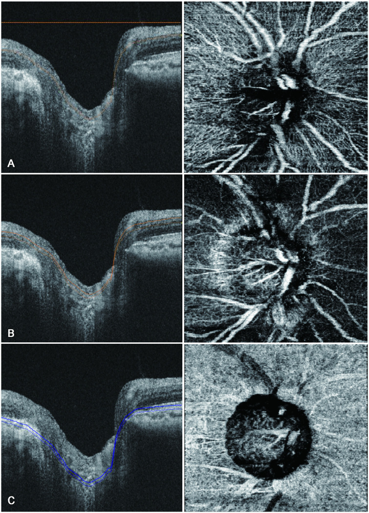
Fig 2. B-scan images of the optic nerve (left) and peripapillary vessels (right) obtained using swept-source optical coherence angiography showing measurement of peripapillary vessel density.
The peripapillary vessel density is measured in the superficial capillary plexus, which comprises the inner capillary layer of the retina (A); the deep capillary plexus, which makes up the outer capillary layer (B); and the choriocapillaris (C).

