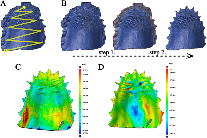
Fig. 1.
The standard scanning pattern of data acquisition was demonstrated on the upper left side (a). The scan was started by a zig-zag movement from the incisive papilla (yellow circle) and finished at the border of the hard and soft palate (yellow box). The preparation of the STL file was demonstrated on the upper right side (b). Teeth were selected (step 1) and removed (step 2) from the scan, and only the palatal area was kept for alignment and surface comparison. The result of the surface comparison with the ISD method of an MZ subject was demonstrated on the lower left side (c), and with ITD method of an MZ twin pair was demonstrated on the lower right side (d). There is one order of magnitude difference between the two scales

