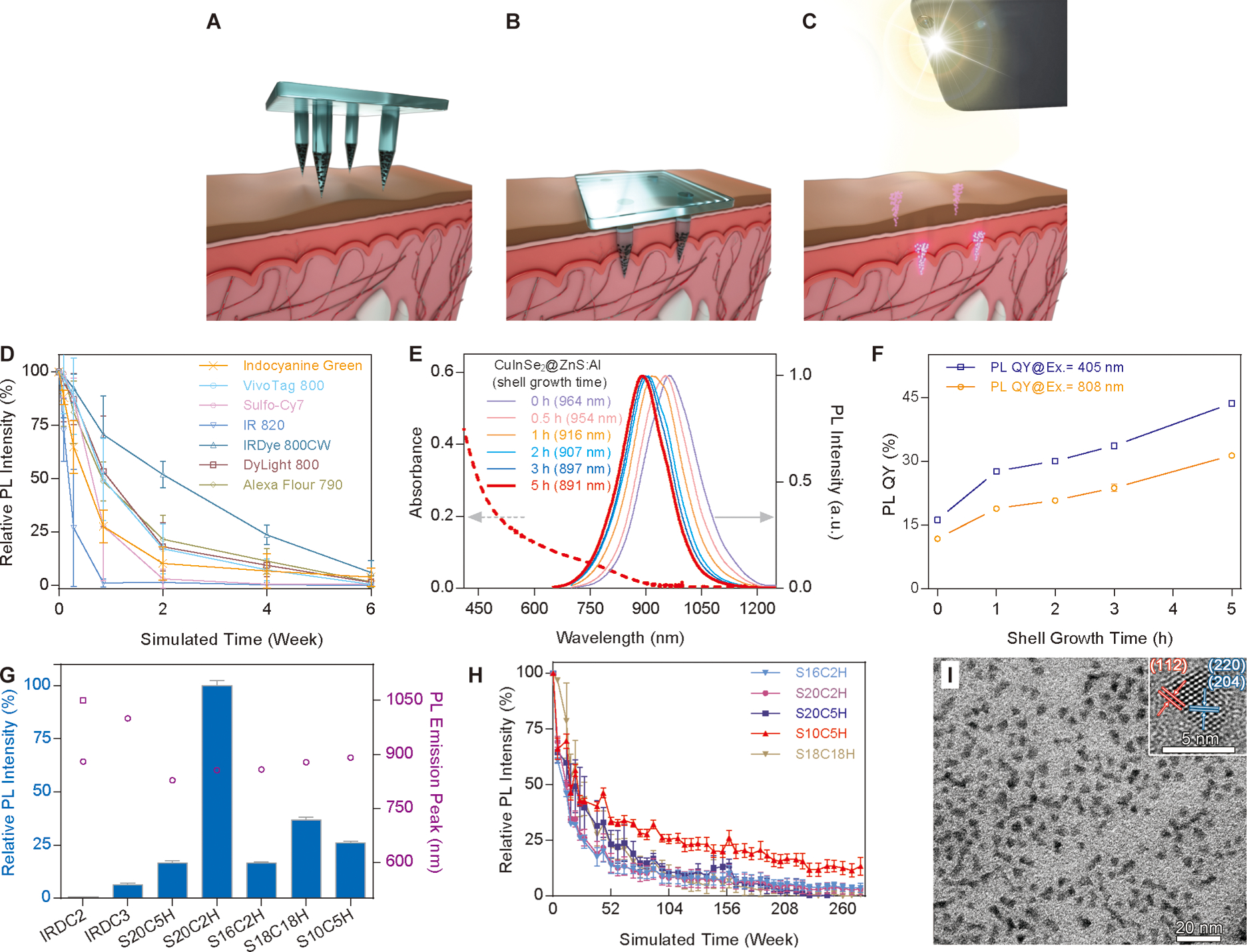
Fig. 1. Platform schematic and fluorescent probe characterization.

(A) Fluorescent microparticles are distributed through an array of dissolvable microneedles in a distinct spatial pattern. (B) Microneedles are then applied to the skin for two to five minutes, resulting in dissolution of the microneedle matrix and retention of fluorescent microparticles. (C) A NIR LED and adapted smartphone are used to image patterns of fluorescent microparticles retained within the skin. By selectively embedding microparticles within microneedles used to deliver a vaccine, the resulting pattern of fluorescence detected in the skin can be used as an on-patient record of an individual’s vaccination history. (D) Rapid photobleaching of organic dyes covered with pigmented human skin under simulated solar light. (E) The emission profiles of QDs (solid lines) show a blue shift with increased shelling time. Dashed line depicts absorption by the 5 h shelling sample. Arrows indicate the relevant y-axis for excitation (left) and emission (right). (F) Photoluminescence quantum yield as a function of shelling time under different excitation wavelengths. (G) Relative photoluminescence intensity comparison of QDs with the commercial inorganic dyes IRDC2 and IRDC3 (blue bars) and corresponding emission peaks (empty purple circles and square). (H) Photostability of QDs covered with pigmented human skin under simulated solar light. (I) Transmission electron microscopy (TEM) and high resolution TEM (inset) showing the size and crystal structure of ZnS:Al-coated CuInSe2 QDs. Scale bars represent 20 nm and 5 nm, respectively. n=3 for all graphs containing error bars, error bars indicate standard deviation.
