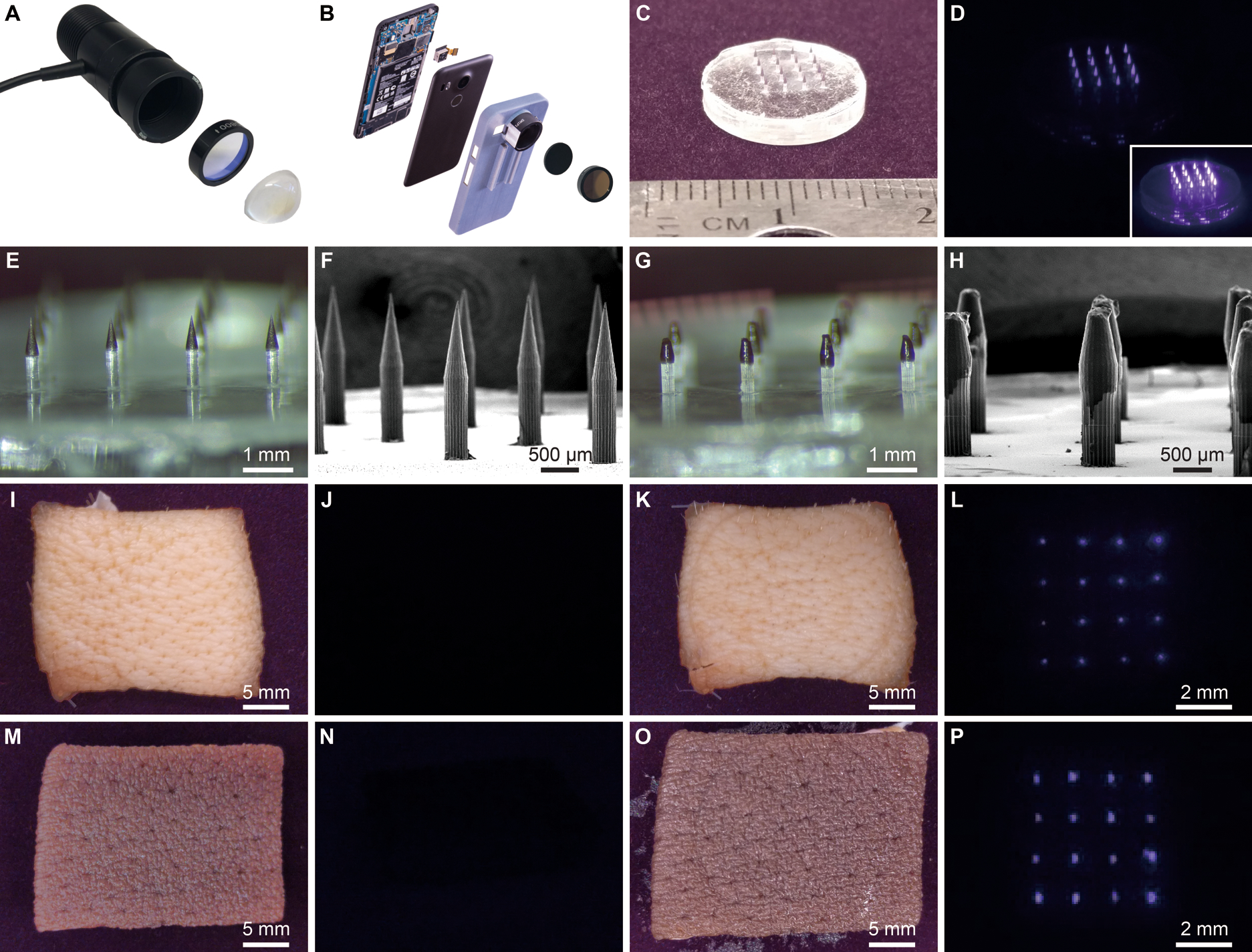
Fig. 4. Smartphone modifications and near-infrared marking detection in skin.

(A) Photograph of disassembled LED used for NIR illumination at 780 nm combined with an 800 nm short-pass filter and aspheric condenser. (B) Photograph of disassembled NIR imaging smartphone consisting of a Google Nexus 5x smartphone with the internal short-pass filter removed and replaced with two external 850 nm long-pass filters set in a 3D-printed phone case. Images of a 16-needle microneedle patch containing PMMA-encapsulated QDs were collected with the adapted smartphone under ambient indoor lighting (C) without the 850 nm long-pass filters and (D) with the pair of 850 nm long-pass filters under LED illumination from the same distance. Inset shows an image at a higher exposure. (E) Optical and (F) SEM images of fluorescent microparticle-loaded microneedles prior to skin application. (G) Optical and (H) SEM images of microneedles after administration to explanted pig skin. Adapted smartphone images of pig skin prior to microneedle application (I) without and (J) with 850 nm long-pass filters. Adapted smartphone images of pig skin after application (K) without and (L) with 850 nm long-pass filters. Adapted smartphone images of pigmented human skin prior to microneedle application (M) without and (N) with the 850 nm long-pass filters. Smartphone images of human skin after application (O) without and (P) with the 850 nm long-pass filters. Note: Scale bars in NIR-filtered images are approximate with (J, L, N, P) taken at about the same distance. Components in panels (A) and (B) cropped for clarity.
