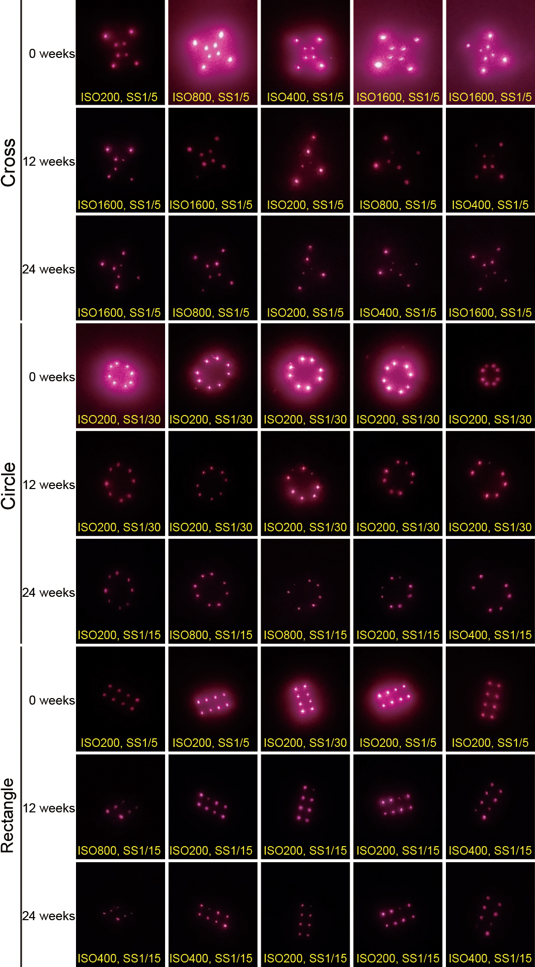
Fig. 6. Longitudinal imaging of NIR markings in rodent skin.

Cropped, but otherwise raw, smartphone images collected from a fixed distance showing the intradermal NIR signal from PMMA-encapsulated QDs delivered via microneedle patches on rats 0, 12, and 24 weeks after administration. The text at the bottom of each image indicates the image collection settings ISO density and shutter speed (SS) in seconds.
