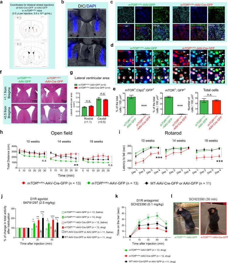
Fig. 1. Effect of striatal mTOR depletion on motor behaviors.
a Schematic representation of the AAV-Cre-GFP or AAV-GFP-injected sites at the indicated coordinates targeting dorsal side of mice striatum. b Representative section showing the DAPI (blue) injection in the striatum using the coordinates in (a). c Confocal images of the striatal brain sections from the mTORflox/flox mice injected with AAV-Cre-GFP or AAV-GFP, showing GFP-Cre or GFP (green) expression, mTOR (blue), and Ctip2 (red) immunohistochemistry, and nuclear stain, DAPI (cyan). d High magnification of confocal images in (c), showing that in AAV-GFP-injected mTORflox/flox mice, Ctip2-positive medium spiny neurons (MSNs) show GFP expression and mTOR immunostaining (yellow arrows). In AAV-Cre-GFP-injected mTORflox/flox mice, Ctip2-positive MSNs express GFP (Cre) but are negative for mTOR immunostaining (white arrows). Some Ctip2-positive MSN negative for GFP (Cre) are positive for mTOR staining (pink arrow). e Quantification for total number of cells identified by DAPI staining, % of mTOR, Ctip2 and GFP triple-positive neurons and % of mTOR and GFP double-positive neurons in striatum of the mTORflox/flox mice injected with AAV-Cre-GFP or AAV-GFP. Images are representative of five ROIs from 4 to 5 sections per animal (n = 4 mice per group). Percentages were determined by considering the number of DAPI stained nuclei as 100%. All values are mean ± SEM. n.s. not significant, ***P < 0.001, two-tailed Student’s t test. f Representative hematoxylin/eosin-stained sections for rostral (+1.1 from bregma) and caudal (+0.5 from bregma) lateral ventricles at the striatal level from the mTORflox/flox mice injected with AAV-Cre-GFP or AAV-GFP. g Quantification of lateral ventricular area from (f). n.s. not significant, two-way ANOVA, Bonferroni post-hoc test (four caudal and four rostral sections were quantified for four mice per group). h, i Total distance (cm) at the indicated time points in open-field test (OFT) (h) and latency to fall (sec) in rotarod test (i) for the mTORflox/flox injected with AAV-GFP (n = 13, female = 10, male = 3), AAV-Cre-GFP (n = 13, female = 6, male = 7) or WT mice injected with AAV-GFP or AAV-Cre-GFP (n = 11, female = 5, male = 6) at 10, 14, and 18 weeks of age. Data are mean ± SEM. **P < 0.01, ***P < 0.001, repeated measures two-way ANOVA followed by Bonferroni post-hoc test. j D1R agonist (SKF81297, 2.5 mg/Kg, i.p.)-induced activity in OFT in AAV-Cre-GFP or AAV-GFP-injected mTORflox/flox and AAV-Cre-GFP/GFP-injected WT mice. Bar graphs indicates % of change in total activity after habituation. Data are mean ± SEM, *P < 0.05, **P < 0.01, ***P < 0.001, repeated measures two-way ANOVA followed by Bonferroni post-hoc test. k Quantification of the catalepsy (time on the bar, sec)-induced by D1R antagonist SCH23390 (0.1 mg/Kg, i.p.) in indicated mice groups. Data are mean ± SEM, n = 11–13 per group, repeated measures two-way ANOVA followed by Bonferroni post-hoc test. l Representative image of catalepsy in AAV-Cre-GFP or AAV-GFP-injected mTORflox/flox mice treated with SCH23390 (30 min). mTORflox/flox injected with AAV-GFP (n = 13, female = 10, male = 3), AAV-Cre-GFP (n = 13, female = 6, male = 7) or WT mice injected with AAV-GFP or AAV-Cre-GFP (n = 11, female = 5, male = 6) were treated with vehicle or drug (j–l).

