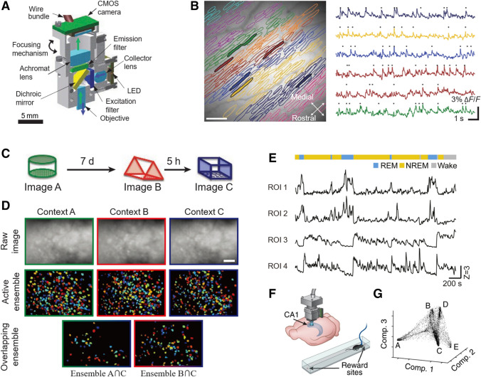
Fig. 1.
Miniature single-photon imaging systems for brain imaging in freely-behaving animals. A Schematic of the integrated headpiece of a wide-field single-photon microscope (LED, light-emitting diode; CMOS, complementary metal-oxide-semiconductor). B Ca2+ spiking dynamics from mouse Purkinje neurons imaged with the system in A (scale bar, 100 μm). Neurons are resolved through computational analysis of their sparse activity amidst a blurred background. Each color denotes one of nine microzones identified. C Experimental design for mice to explore three different contexts (A, B, and C), each for 5 or 10 min separated by either 5 h or 7 days. D CA1 neuronal activity during context exploration according to C. Upper, images of mean fluorescence from each session; middle, ensemble of cells active in each session; lower, cells active in two sessions (scale bar, 100 μm). E Activity of galaninergic neurons in the dorsomedial hypothalamus during sleep. Upper, brain states (color coded); lower, Ca2+ traces (Z scores) from four regions of interest (ROI) in the same field of view. REM, rapid eye movement; NREM, non-REM. F Schematic of the experimental setup. Imaging via a miniature single-photon microscope combined with a GRIN lens. Mice run back and forth on a linear track. G Data of CA1 neuronal activity viewed in the reduced dimensional space of three components, which are the three leading eigenvectors, based on the non-linear dimensionality reduction algorithm Laplacian Eigenmaps. A, B are from [22], C, D from [29], E from [33], F, G from [34].

