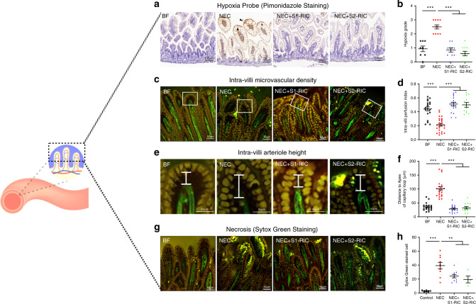
Fig. 5. RIC improves intestinal villi microvasculature and reduces ischemia and necrosis of enterocytes at the villi tip.
a, b Pups were injected with pimonidazole, a sensitive marker which allows localization of intestinal ischemia. Data represent immunohistochemistry of pimonidazole in the ileum comparing breastfed (BF) control (n = 8), NEC (n = 10), NEC with Stage 1 RIC (n = 10), and NEC with Stage 2 RIC (n = 10). Scale bars are equivalent to 100 µm in the images shown. c Intra-villi microvasculature was investigated in RosamT/mG/+;Tie2-Cre using TPLSM and compared between BF, NEC, NEC with Stage 1 RIC, and NEC with Stage 2 RIC (n = 4 per group; and minimum of 2 measurements were obtained per group). d Villus perfusion index was calculated as the ratio of the area of intra-villi arterioles to the area of the whole villi and compared between the listed groups. e, f The distance between the apex of the capillary loop and the apical side of the villi epithelium was measured and compared between listed groups. g Cell death (yellow staining in the tip of villi) in the ileal epithelium was detected using Sytox Green, a marker of necrosis which does not permeate into live cells but binds to cellular nucleic acids in dead cells, staining them with intense green fluorescence and was compared between listed groups. h Necrotic cells were counted in the listed groups (*p < 0.05; **p < 0.01; ****p < 0.0001). Data were compared using two-sided one-way ANOVA with post hoc Turkey test (**p < 0.01; ****p < 0.0001). Scale bars in (c, e and g are equivalent to 50 µm, and data are presented as mean ± SEM. Source data are provided as a Source Data file.

