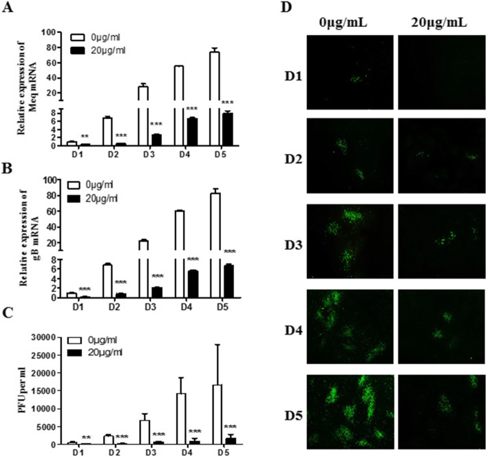
Fig. 3.
Time-dependent manner of baicalin inhibition on MDV replication in CEF. Total cellular RNA was extracted from day 1 to day 5 p.i., and the expression levels of Meq gene (a) and gB gene (b) were detected by qRT-PCR. c The results of plaque count of the MDV RB-1B strain infected CEF treated with 0 and 20 μg/mL of baicalin at different time points. d Direct observation of viral plaque formation dynamics by indirect immunofluorescence assays from day 1 to day 5 post virus infection. Data were expressed as mean ± SD is for A, B and C only from three independent experiments and analyzed by Student’s t-tests (**p < 0.01, *** p < 0.001)

