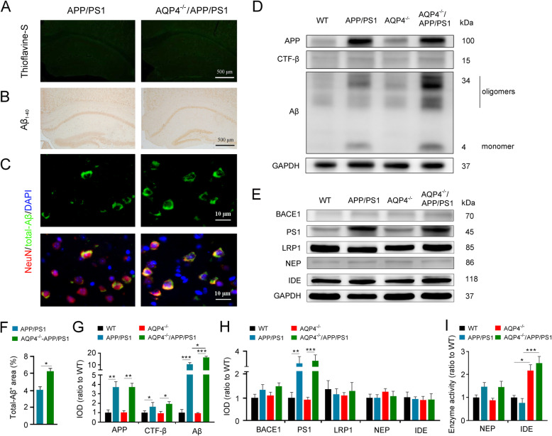
Fig. 2.
AQP4 deletion increased intraneuronal Aβ and IDE activity without causing plaque formation in the cerebral cortex of 3-month-old APP/PS1 mice. a, b Brain sections stained by thioflavine-S florescence and Aβ1–40 immunohistochemistry showing no Aβ plaque disposition. c Double immunofluorescence for total-Aβ and NeuN. There was increased Aβ immunostaining in cerebral neurons of AQP4−/−APP/PS1 mice compared to those in APP/PS1 controls. d, g Representative Western blot bands and densitometry analysis of APP, CTF-β, and Aβ monomer/oligomers. e, h Representative Western blot bands and densitometry analysis of BACE1, PS1, LRP1, IDE, and NEP in the cortex. f Quantification of Aβ-positive area fraction. i Enzyme activity assay of NEP and IDE. Data in f were analyzed by Student’s t test; other data were analyzed by the two-way ANOVA with Tukey’s post hoc test. Data are mean ± SEM. n = 4 per group. *p < 0.05; **p < 0.01; ***p < 0.001

