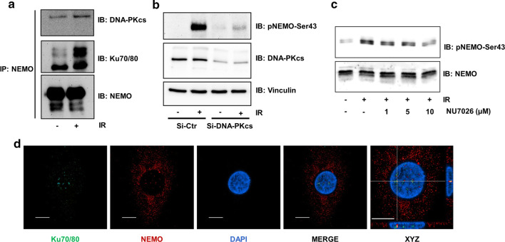
Fig. 4.
DNA-PK/IKKγ interaction in primary macrophages. a CoIP of NEMO and DNA-PK subunits from the lysates of macrophages after exposure of cells to irradiation (IR, 5 Gy) and subsequent incubation for 1 h. b Human primary macrophages were transfected with either GL3 control siRNA (siCtr) or siRNA targeting the DNA-PK subunit (siDNA-PKcs). After pretreatment with siRNA for 24 h, cells were exposed to IR (5 Gy), and lysates were analysed by immunoblotting (IB) with specific antibodies as indicated. c Human primary macrophages were incubated in the presence of different doses of the DNA-PK inhibitor NU7026 for 2 h. After exposure to irradiation (IR, 5 Gy), lysates were analysed by IB with specific antibodies as indicated. d Immunofluorescence staining of macrophages with antibodies against Ku 70/80 (green) and NEMO (red) and nuclear staining with DAPI (blue). In the merged image, the magnified region of interest (right picture) and particularly the xz and yz projections of individual clusters of Ku 70/80 and NEMO were localized very close to each other in the euchromatin of the nucleus. Scale bar indicates 10 µm

