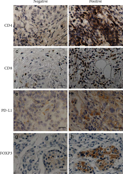
Figure 1.

Immunohistochemical staining: (a) negative CD4+ T-cell infiltration and (b) positive CD4+ T-cell infiltration; (c) negative CD8+ T-cell infiltration and (d) positive CD8+ T-cell infiltration; (e) negative PD-L1 expression and (f) positive PD-L1 expression; (g) negative FOXP3+ Treg cell infiltration and (h) positive FOXP3+ Treg cell infiltration. Scale bars represent 50 μm.
