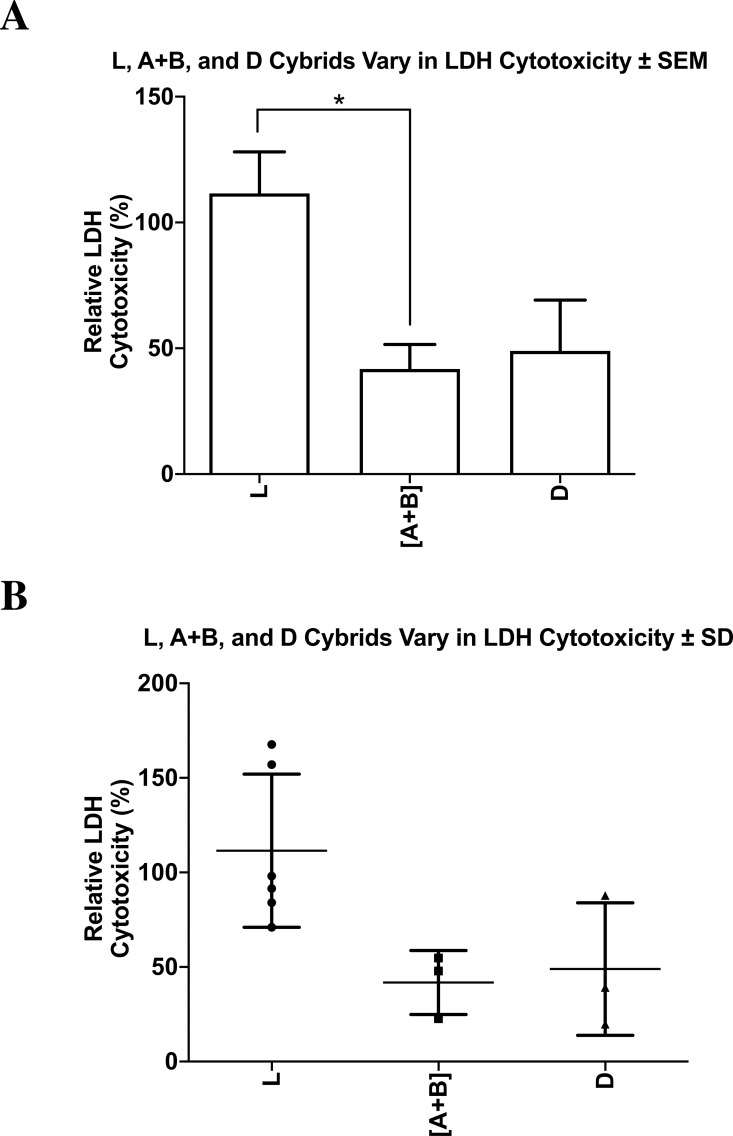
Figure 9. L, [A+B], and D cybrids vary in LDH cytotoxicity.
The cytotoxicity levels of L, [A+B], and D cybrids were measured with the lactate dehydrogenase (LDH) assay. Cybrids cultured to the fifth passage were plated in 96-well plates (10,000 cells/ well) for 24 h and treated with 0 or 40µM of cisplatin for another 48 h. The LDH assay was conducted and absorbance values were taken at both 490 nm and 680 nm using an absorbance reader. Percent cytotoxicity levels were calculated using the equation (Treated LDH level –spontaneous LDH level) divided by (Maximum LDH level –spontaneous LDH level) multiplied by one hundred. (A) All values were normalized to the average of the untreated-L cybrids and displayed as the mean ± Standard Error Margin (SEM). (B) The distribution of each value was also presented in a dot-plot graph representing mean ± Standard Deviation (SD). When normalized to the L cybrids, there were significantly higher LDH cytotoxicity levels in L cybrids compared to [A+B] cybrids (111.5% ± 16.5% SEM; SD = 40.5%, n = 6 versus 41.8% ± 9.8% SEM; SD = 16.9%, n = 3, respectively) (p = 0.0270). The D cybrids trended lower LDH levels but they were not significant (48.9% ± 20.2% SEM; SD = 35.0%, n = 3, p = 0.0576) compared to the L cybrids. LDH cytotoxicity levels between the [A+B] and D cybrids were not statistically different (p = 0.766). The entire experiment was repeated three separate times. ∗p < 0.05; ∗∗p < 0.01; ∗∗∗p < 0.001.

