Figure 3:

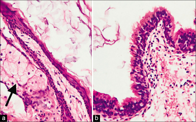
High power magnification showed (a) pilosebaceous units (arrow) embedded in cyst wall and (b) focal lining by stratified ciliated columnar epithelium. (H&E, 400×).

High power magnification showed (a) pilosebaceous units (arrow) embedded in cyst wall and (b) focal lining by stratified ciliated columnar epithelium. (H&E, 400×).