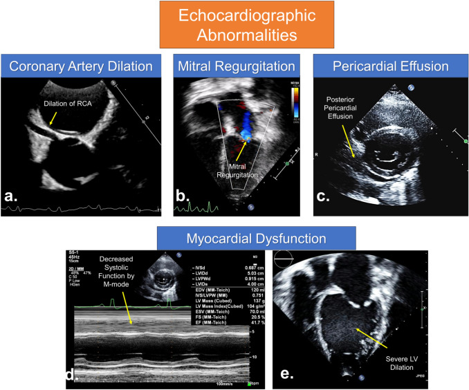
Fig. 3.
Echocardiographic abnormalities in children with COVID-19 and MIS-C. a Parasternal short axis view at the level of aortic valve demonstrating diffuse dilation of right coronary artery (RCA) and coronary artery walls are echogenic. b Apical 4 chamber view demonstrating mitral valve regurgitation. c Parasternal short axis view at the level of mitral valve showing tiny posterior pericardial effusion in a patient with myocarditis. d M-mode demonstrating decreased systolic function. e Apical 4 chamber view demonstrating severe left ventricular (LV) dilation due to myocardial dysfunction

