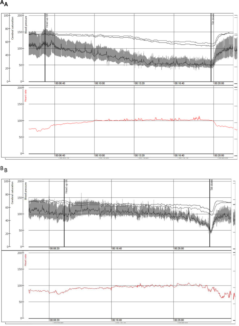
Figure 2. Classical and Delayed Orthostatic Hypotension on Tilt.

shows in panel A: Beat to beat blood pressure (mmHg) from a photoplethysmographic finger device and heart rate (beats/min) in upper section with cerebral saturation (%) from a near-infra-red spectrum device in the lower section during head-up tilt in a female patient aged 50 years with classical orthostatic hypotension. Panel B shows, in a similar format to Panel A: Delayed orthostatic hypotension leading ultimately to vasovagal reflex in a male patient of 80 years. Reproduced with permission of Frontiers Cardiovascular Medicine.
