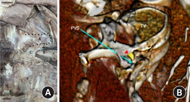
Fig. 2.

Paravertebral anatomy. (A) Landmarks of the paravertebral space (PVS) are shown in deep dissection and (B) with corresponding reconstructed computed tomography angiography using Anatomage system. A: thoracic transverse process, B: intercostal neurovascular bundle, C: partially dissected superior costotransverse ligament, D: pleura, red arrowhead: spinal nerve, PVS at tip of blue arrow: paravertebral space, R: rib, SP: spinous process, TP: transverse process. Notice the location of the neurovascular bundle immediately below the TP in the dissection.
