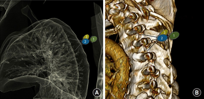
Fig. 5.

Relationship between lung and potential needle placement. (A) Anatomic reconstruction of lung windows (B) with reconstructed computed tomography of thoracic spine in sagittal plane. Numbers indicate 1: needle tip location with initial placement, 2: cephalad reorientation & 1.5 cm advancement, and 3: caudal reorientation & 1.5 cm advancement. Note proximity of the needle tips to the lung parenchyma with either direction.
