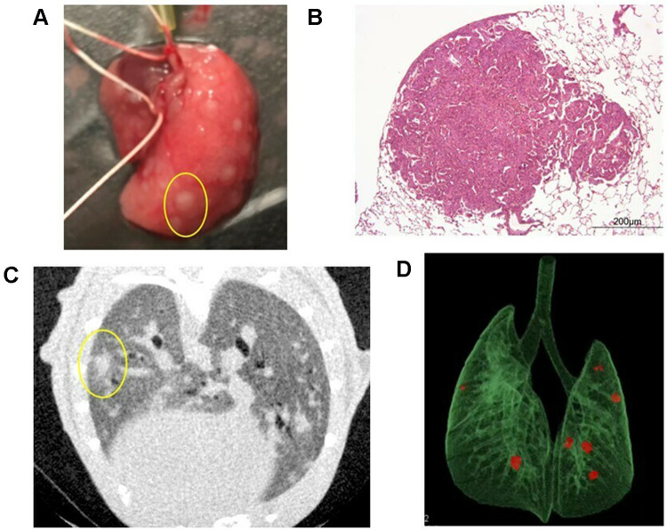
Figure 2.
Pulmonary metastases. (A) Macroscopic findings of pulmonary metastasis in the right lobe of the lung. In this mouse, many nodules were identified (one is circled in yellow). (B) Hematoxylin-eosin staining of pulmonary metastasis in a mouse. (C) A representative two-dimensional image of a micro CT scan in a smoking mouse. In this mouse, one nodule was identified in the left lung (circled in yellow). (D) CT reconstruction of a pulmonary metastasis from a mouse model. Three-dimensional microstructural image data were reconstructed using Tri/3D-BON software. Red colored nodules indicate pulmonary metastasis.

