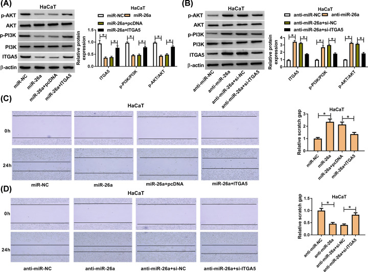
Figure 5. ITGA5 reversed the effect of miR-26a on cell migration via PI3K/AKT signaling pathway.
(A) The expression level of ITGA5 was detected by Western blot assay after transfection with miR-26a or miR-26a+ITGA5 in HaCaT cells. (B) The expression level of ITGA5, PI3K and AKT was measured after transfection with anti-miR-26a or anti-miR-26a+si-ITGA5. (C,D) The migration ability of HaCaT cell was analyzed after overexpressing both miR-26a and ITGA5 in HaCaT cells after TGF-β1 treatment for 24 h. (B) The migration cell number of HaCaT after transfection of miR-26a inhibitor and si-ITGA5 after TGF-β1 treatment for 24 h. * means P-value less than 0.05.

