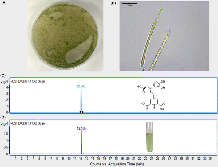
Fig. 2.

Detection of dopaxanthin in cultures of Anabaena cylindrica supplemented with L‐DOPA.
A. 3‐day plate of BG‐11 medium containing A. cylindrica.
B. Microscopy image of the filamentous distribution of A. cylindrica cells. Scale bar: 20 μM.
C and D. Chromatograms from HPLC‐ESI‐TOF‐MS analysis. Standard dopaxanthin was followed at EIC 391.1136 m/z (C) and the same peak was detected in culture media of A. cylindrica supplemented with L‐DOPA 7.6 mM, 15 mM sodium ascorbate and 100 mM EDTA (D).
