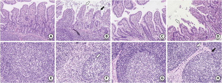
Fig. 1. Morphological changes within the ileum of piglets during PCV2 infection. (A) Histomorphologic observations of the ileum either without PCV2 infection (21 dpi, 100×) or (B) after PCV2 infection (21 dpi, 100×). As indicated by the white arrow, the intestinal epithelial cells of the villus fall off, and the black arrow shows shortening of the villus in the infection group. The mucosal tissues in the infection group were markedly infiltrated with inflammatory cells. (C, D) At 56 dpi, as indicated by the white arrow, the shape and size of the villi were partially restored. On the other hand, the mucosal tissues were still infiltrated with inflammatory cells in the infection group (56 dpi, 100×). (E) The mesenteric lymph nodes of the control group (21 dpi, 200×). (F) The mesenteric lymph nodes of the infected group (21 dpi, 200×). The white arrow indicates that there are fewer lymphocytes. (G) The mesenteric lymph nodes of the control group (56 dpi, 200×). (H) The mesenteric lymph nodes of the infection group (56 dpi, 200×). Scale bar, A-D: 200 μm, E-H: 100 μm.
PCV2, porcine circovirus type 2; dpi, days post-infection.

