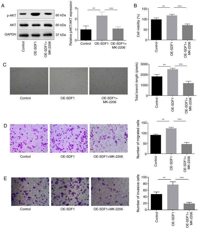
Figure 4.
Influence of AKT inhibition on the angiogenesis of VECs. VECs with MK-2206 (10 µM, 30 min) treatment or non-treated were cultured in the cultural supernatant derived from NPCs with OE-SDF1 or controls, and then the following assays were performed. (A) Western blotting was performed to detect the expression levels of p-AKT and AKT in VECs. (B) VEC viability was detected by Cell Counting Kit-8. (C) Tube formation ability in VECs was analyzed by Matrigel tube formation assay, and the total branch length was calculated by ImageJ (magnification, ×100). (D and E) Migration and invasion abilities of VECs were analyzed by Transwell assay, with SDF1 overexpressed or non-treated NPCs as a chemokine (magnification, ×100). n=3, **P<0.01, ***P<0.001. SDF1, stromal cell-derived factor 1; NPCs, nucleus pulposus cells; OE-, overexpression; p-, phosphorylated; AKT, AKT serine/threonine kinase 1; VECs, vascular endothelial cells.

