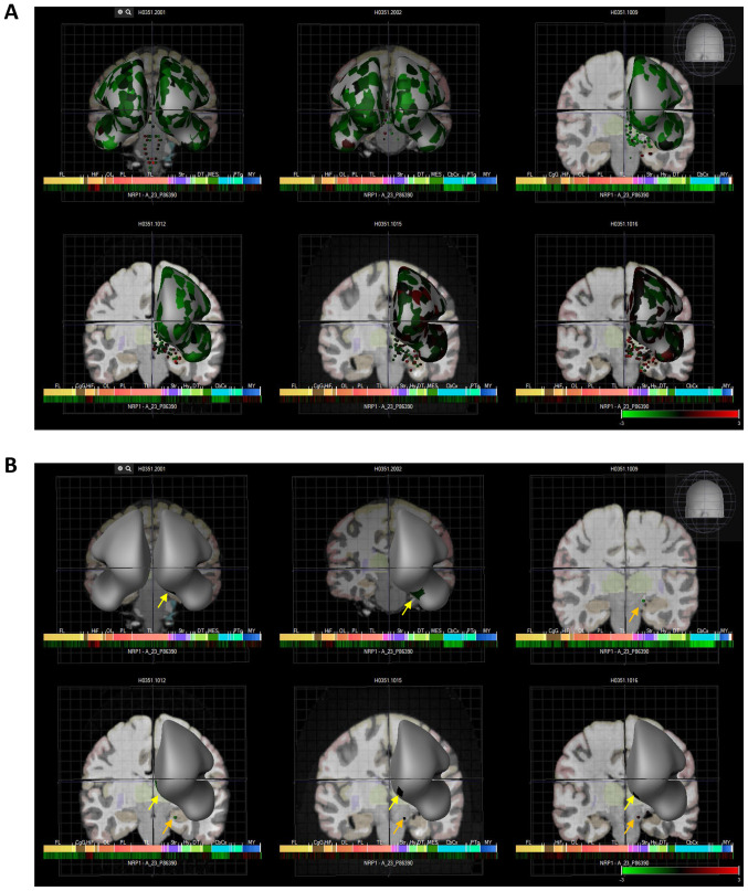
Figure 3.
Six human donors and heat map expression of NRP1 as assessed by microarray (Allen brain atlas). (A) Indicates global brain expression of NRP1 as detected by microarray probes, with expression of cerebral areas indicated on the hemispheres and expression in brain nuclei indicated as dots. Heat maps of expression are displayed below individual brains and organised by anterior to posterior regions with the FL, HiF, OL, PL, TL, Str, DT, CbCx and MY marked on the heat map. (B) Shows expression in olfactory accessory tissues with yellow arrows indicating the paraolfactory gyri and orange arrows indicating the olfactory tubercle. NRP1, neuropilin-1; FL, frontal lobe; HiF, hippocampal formation; OL, occipital lobe; PL, parietal lobe; TL, temporal lobe; Str, striatum; DT, dorsal thalamus; CbCx, cerebral cortex, MY, myelencephalon.

