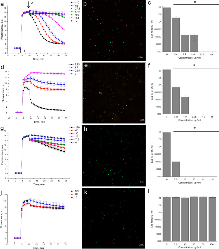
FIGURE 3.
Evaluation of membrane disturbance of S. aureus 209P using Live/Dead fluorescent dyes and agar-drop assay. (a,d,g,j) Intensity of SYTO 9 fluorescence. If bacterial membranes are permeabilized, propidium iodide penetrates into the cell. What follows is SYTO 9 getting displaced from nucleic acids, which leads to a decrease in fluorescence intensity in the green region (505–540 nm) of the spectrum. (b,e,h,k) Epifluorescence microscopy images of S. aureus 209P after 1 h of treatment. (c,f,i,l) Cell’s viability after exposure with antibiotics for 1 h. 5812-A/C (a–c), nisin (d–f), daptomycin (g–i), vancomycin (j–l). 1—The moment of introduction of the fluorophore; 2—the moment of introduction of the antibiotic. *Indicate significant differences at p < 0.05.

