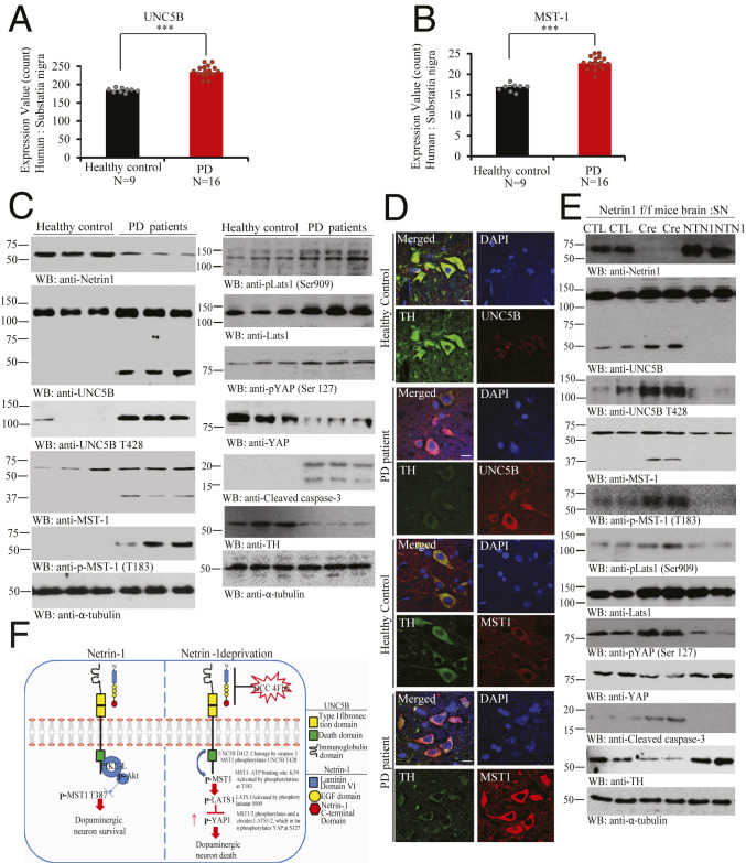
Fig. 7.
UNC5B and MST1 phosphorylation and activation in PD patient’s brains. (A and B) Quantification of UNC5B and MST1 expression levels in healthy control or PD patient brains. (A) UNC5B and (B) MST1 genes expression profiling by arrays of PD SN from datasets GDS2821. (Unpaired t test, ***P < 0.0005, means + SD are shown.) (C) NTN1 reduction in PD brains is associated with Mst1 activation and phosphorylation of UNC5B and TH loss. Immunoblot of NTN1, UNC5B, p-UNC5B (T428), MST1, p-MST1 (T183), p-LATS1 (Ser909), LATS1, p-YAP (Ser127), YAP, TH, and cleaved caspase-3 levels in age-matched healthy control vs. PD patients brain tissue samples. Three independent studies were conducted in all of the experiments. (D) IF costaining reflects the inverse correlation of MST1/TH and UNC5B/TH in the human brain sections. The SN regions from healthy control or PD patient brain sections were costained with indicated antibodies. TH/UNC5B or TH/MST1 coimmunostaining representative images were conducted in age-matched healthy control vs. PD patient’s brain paraffin slides. Three independent blinder tests were conducted in all of experiments. (Scale bar: 50 µm.) (E) NTN1 depletion from NTN1 f/f mice elicits MST1 phosphorylation of UNC5B T428 and TH loss, which is blocked by NTN1 overexpression. Representative immunoblotting images of SN tissues from AAV6-GFP (control virus), AAV6-GFP Cre, or hNTN1 virus-injected NTN1 f/f mice brain. Three independent studies were conducted in all of the experiments. (F) The schematic model of NTN1 deprivation-elicited Hippo/MST1 signaling pathway via UNC5B receptor. CTL, control.

