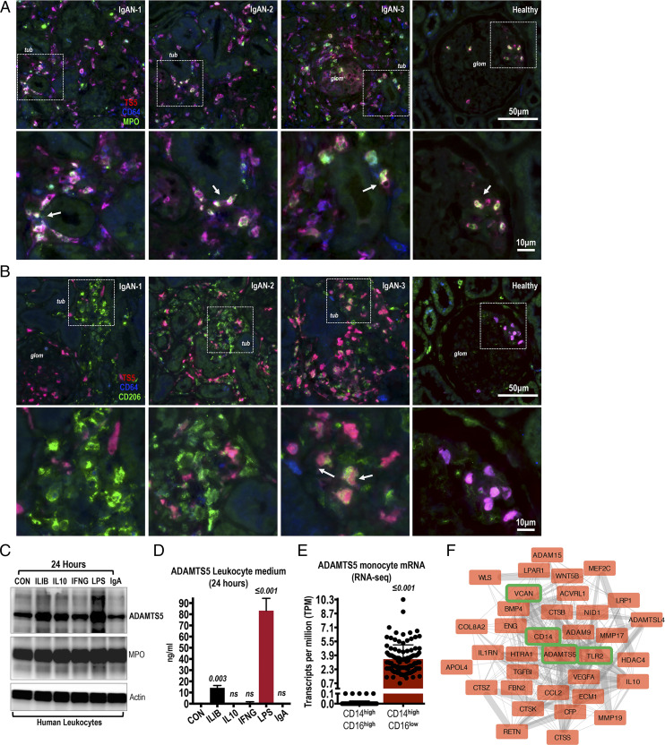
FIGURE 2.
Costaining of ADAMTS5 in IgAN biopsies with inflammatory cell markers. (A) In IgAN and healthy kidney biopsies, ADAMTS5 (red) was costained with CD64 (blue) and myeloperoxidase (MPO; green). ADAMTS5 colocalises with CD64+ cells in both IgAN and healthy biopsies (cells with ADAMTS5 and CD64 colocalisation appear as purple). There are also MPO+ cells and sporadic triple-positive ADAMTS5-CD64-MPO (white) cells were present indicating expression of ADAMST5 by infiltrating monocytes and neutrophils (see arrows). Note intense accumulation of ADAMTS5+-CD64+-MPO+ cells in the interstitium around tubules (tub) in affected IgAN specimens. In healthy biopsies, triple-stained cells were mainly seen in glomeruli (glom), presumably naturally perfusing circulating leukocytes. (B) ADAMTS5 (red) was costained with CD206 (green) and CD64 (blue). ADAMTS5 colocalises with CD64 (purple cells). In contrast, colocalisation of ADAMTS5 (red) with interstitial CD206 (green) is weak and sporadic in inflamed areas (see IgAN-3; arrows) indicating different cellular origins. (C) ADAMTS5 was immunoblotted on leukocyte lysates. Leukocytes were isolated from healthy donors by erythrocyte lysis and were cultured for 24 h with 100 ng/ml rIL-1B, IL-10, IFNG, 1 μg/ml LPS, or 200 μg/ml IgAN-serum jacalin-purified IgA. Myeloperoxidase was used for comparison and βActin as a loading control. (D) ADAMTS5 protein levels measured by ELISA in the culture medium of leukocytes stimulated for 24 h as in (C). (E) RNA sequencing expression of ADAMTS5 in classical versus nonclassical monocytes. Data retrieved from the Human Protein Atlas and (26). (F) ADAMTS5 first-degree neighbors (proteins interacting directly with ADAMTS5 via a single edge) derived from the classical monocyte elevated gene interactome. Classical monocyte elevated genes were downloaded from the Human Protein Atlas. Protein-protein interactions were processed and visualized in Cytoscape.

