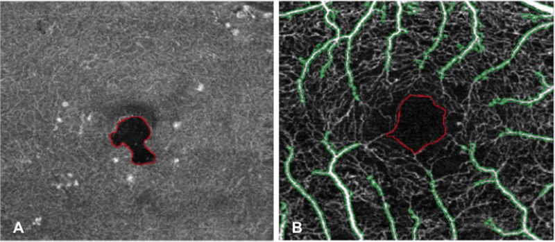
Figure 1.
MATLAB (version 2017b, Mathworks, Natick, MA) analyses performed on OCTA images of the same patient (left eye) to evaluate macular perfusion parameters. (A) First method of analysis: automatic detection and demarcation of cyst area in the en-face DCP image; the area of the cyst was then excluded from final computation of perfusion parameters. (B) Second method of analysis: OCTA image of the full macula slab in which the area corresponding to FAZ (identified by red boundaries) and noncapillary blood vessels (identified by green boundaries) was excluded from final computation of PCD. OCTA, optical coherence tomography angiography; DCP, deep capillary plexus; FAZ, foveal avascular zone; PCD, perfused capillary density.

