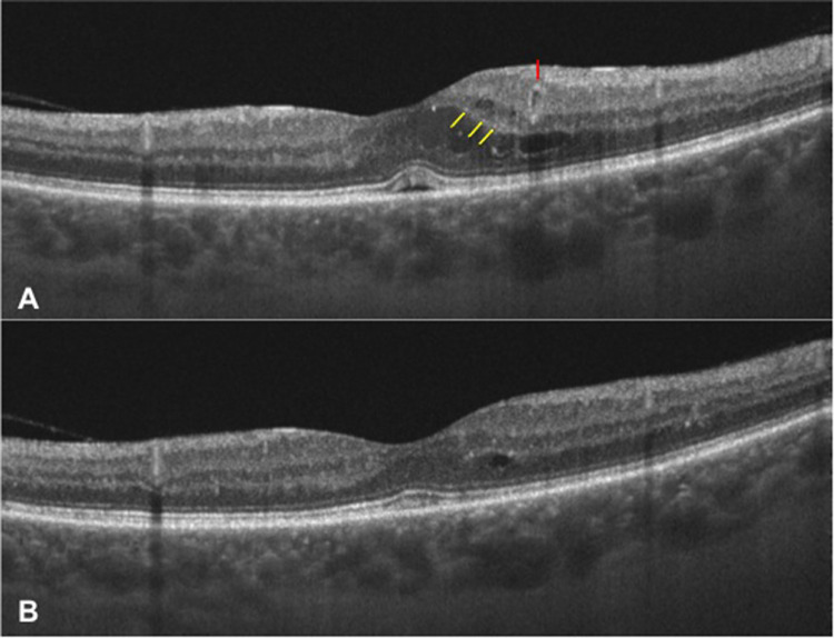
Figure 5.
Images of the left eye of a patient with DME at baseline (A) and 12 months after SMPL treatment (B) showing the reduction of HRS (just a few of the HRS highlighted by yellow arrows at baseline), complete resolution of serous retinal detachment and disappearance of a perifoveal microaneurysm (highlighted by a red arrow at baseline). At the 12-month visit, all retinal layers are clearly discernible and no DRIL is present. DME, diabetic macular edema; SMPL, subthreshold micropulse laser; HRS, hyper-reflective retinal spots; DRIL, disorganization of inner retinal layers.

