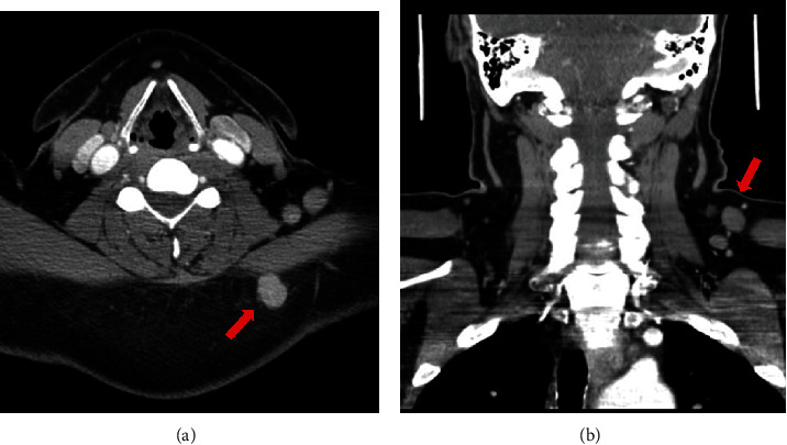
Figure 1.

(a, axial view) CT scan with contrast demonstrated a hyperdense rounded mass in the subcutaneous tissue of the left side of the neck, posterior to the trapezius muscle, corresponding to the patient's palpable primary mass (red arrow). (b, coronal view) Several lymph nodes (red arrow) in the posterior triangle anterior to the trapezius muscle ranging in size up to 1.5 cm in diameter were demonstrated on CT scan.
