Fig. 3.
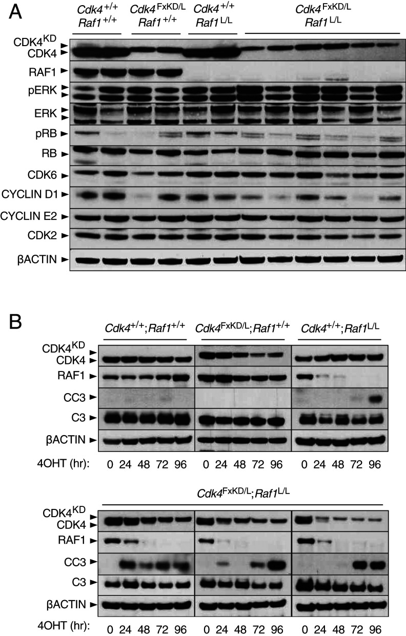
Concomitant CDK4 inactivation and RAF1 ablation boosts apoptosis. (A) Western blot analysis of CDK4, CDK4KD, RAF1, phospho-ERK (pERK), ERK1/2 (ERK), phospho-RB (pRB), RB, CDK6, CYCLIN D1, CYCLIN E2, and CDK2 expression in lysates obtained from Kras+/G12V;Trp53−/−;hUBC-CreERT2+/T lung tumor cell lines harboring Cdk4+/+;Raf1+/+, Cdk4FxKD/L;Raf1+/+, Cdk4+/+;Raf1L/L, and Cdk4FxKD/L;Raf1L/L alleles 96 h after 4OHT exposure. β-Actin was used as loading control. Migration of the above proteins is indicated by arrowheads. (B) Western blot analysis of CDK4, CDK4KD, RAF1, cleaved caspase-3 (CC3), caspase-3 (C3) expression in lysates obtained from Kras+/G12V;Trp53−/−;hUBC-CreERT2+/T lung tumor cell lines harboring Cdk4+/+;Raf1+/+, Cdk4FxKD/L;Raf1+/+, Cdk4+/+;Raf1L/L, and Cdk4FxKD/L;Raf1L/L alleles maintained in 4OHT containing media. Samples were harvested at the indicated times (hr, hours). β-Actin was used as loading control. Migration of the above proteins is indicated by arrowheads.

