Fig. 5.
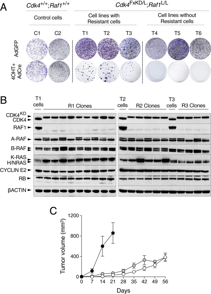
Generation of CDK4/RAF1 resistant tumor cell clones. (A) Kras+/G12V;Trp53−/−;hUBC-CreERT2+/T;Cdk4+/+;Raf1+/+ (C1 and C2) and Kras+/G12V;Trp53−/−;hUBC-CreERT2+/T;Cdk4FxKD/L;Raf1L/L (T1 to T6) lung tumor cell lines exposed to 4OHT and infected with AdCre to inactivate CDK4 and RAF1 were used in a colony formation assay to identify resistant clones. Adeno GFP (AdGFP) was used as negative control. (B) Western blot analysis of CDK4, CDK4KD, RAF1, A-RAF, B-RAF, K-RAS, H-RAS, N-RAS, CYCLIN E2, and RB expression levels in lysates of parental T1, T2, and T3 cell lines expressing CDK4 and RAF1 and of resistant clones (R1, R2, and R3 clones) derived from these cell lines upon CDK4 inactivation and RAF1 ablation. β-Actin was used as loading control. Migration of the above proteins is indicated by arrowheads. (C) Tumor growth of individual CDK4/RAF1 resistant clones R1.1, R1.2, and R1.3 (solid circles), R2.1, R2.2, and R2.3 (open circles), and R3.1, R3.2 and R3.3 (gray circles) after subcutaneous implantation in immunodeficient mice. Error bars indicate mean ± SEM.

