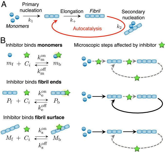
Fig. 1.
Microscopic mechanisms of protein aggregation and possible inhibition pathways. (A) Schematic representation of the microscopic steps of protein aggregation into fibrillar structures. (B, Left) Potential target species during protein aggregation and associated binding rate constants. (B, Right) Schematic diagrams showing the microscopic steps that are targeted by the inhibitor.

