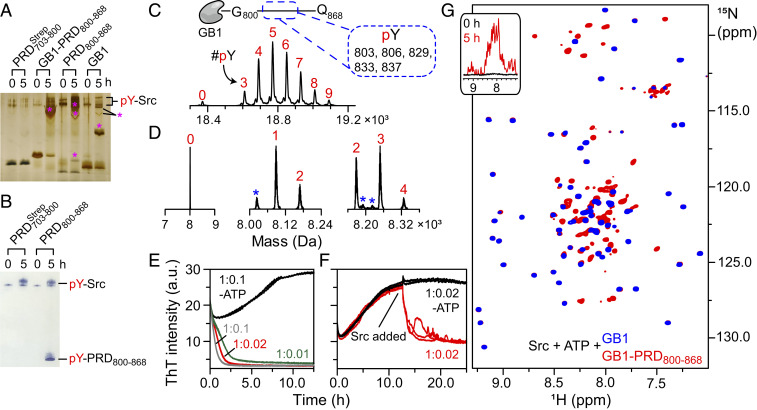
Fig. 7.
Dissolution of GB1–PRD800–868 amyloids upon Src-mediated tyrosine phosphorylation. (A–D) Characterization of in vitro phosphorylation of PRD constructs using Phos-tag SDS-PAGE (A), Western blotting (B), and MS (C and D). For Phos-tag gel, the following constructs, namely GB1–PRD800–868, PRD800–868, and the GB1 tag, were incubated with Src (substrate to kinase molar ratio: 1:0.01). Phosphorylated products were visualized by silver staining and are marked by pink asterisks. (B) In vitro phosphorylation of and PRD800–868 by Western blotting. (C and D) LC-ESI-TOFMS and LC-MS/MS analyses of in vitro phosphorylation reactions revealed hyperphosphorylated states of GB1–PRD800–868 (C) and PRD800–868 (D). A schematic representation of GB1–PRD800–868 along with phosphorylated tyrosines (dashed rectangle) are shown above the graph in C. The numbers in red represent the number of phosphorylated tyrosines, labeled as pY (peaks marked with blue asterisks represent sodium/iron adducts; SI Appendix, Table S2). (E–G) The impact of tyrosine phosphorylation on aggregation kinetics of GB1–PRD800–868 was assessed using ThT assays (E and F) and NMR spectroscopy (G). For ThT assays (n = 3) (E), 150 µM GB1–PRD800–868 samples were incubated at 30 °C with 2 mM ATP and varying concentrations of Src (molar ratios: 1:0.1 [gray], 1:0.02 [red], and 1:0.01 [green]). a.u., arbitrary units. Control experiments were carried out on a GB1–PRD800–868 + Src mixture in the absence of ATP (black; molar ratio: 1:0.1). (F) Samples of 150 µM GB1–PRD800–868 were incubated at 30 °C without Src (n = 5) for ∼11 h. Src + ATP were then added to three samples (red), whereas the remaining two (black) received only Src. G shows the overlay of expanded regions of the 1H-15N TROSY-HSQC spectra of phosphorylated GB1–PRD800–868 (red) and the phosphorylated GB1 tag (blue). Both were incubated with Src (molar ratio: 1:0.1) in the presence of 2 mM ATP for 5 h at 30 °C. (G, Inset) Corresponding one-dimensional profiles of 15N-labeled GB1–PRD800–868 recorded at 0 and 5 h (black and red, respectively) after addition of unlabeled Src and ATP.

