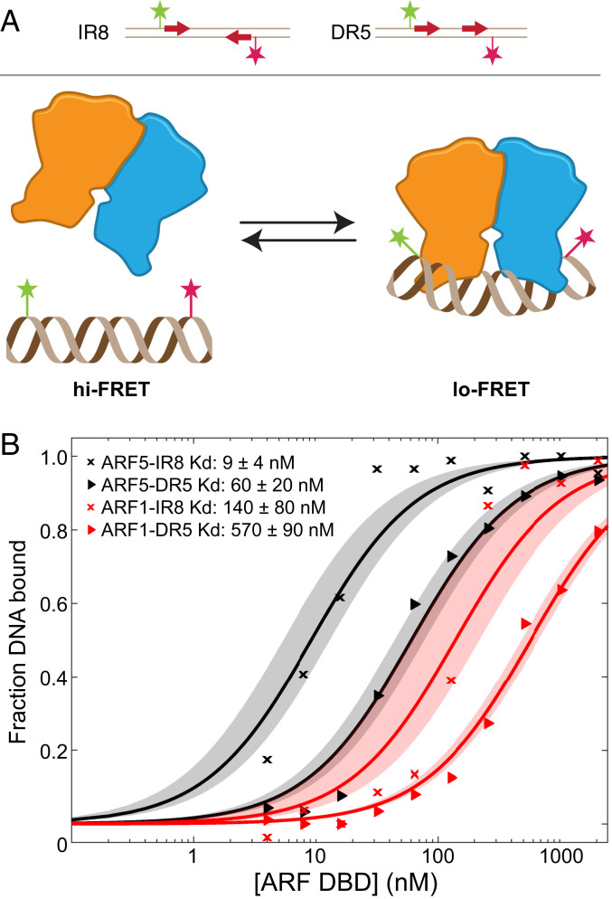
Fig. 6.
Single-molecule FRET assays reveal differential ARF-DNA affinities. (A) Cartoon describing the single-molecule FRET assay. IR8- or DR5-containing (arrows) ds oligonucleotide carrying two FRET-compatible dyes (magenta and green stars) are immobilized on a coverslip. Arrows indicate position of TGTCGG sequences. Binding of ARF proteins (dimer indicated in orange/blue) to the oligo leads to bending of DNA bending and slight displacement of the dyes thereby decreasing FRET efficiency. (B) Titrations of ARF1-DBD and ARF5-DBD proteins (concentration in nM on x-axis) on surface-immobilized DR5 or IR8 oligonucleotides. The y-axis shows the fraction of DNA bound to protein derived from FRET efficiency distribution. Dissociation constants (Kd) are given in the legend, with their 95% intervals of confidence from the fit.

