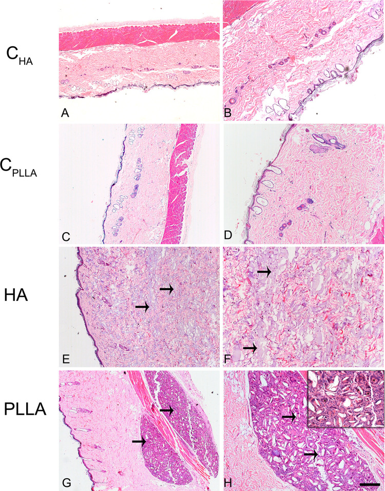
Figure 4.
Photomicrograph of dermal skin biopsies after 60 days of dermal injection of PBS (control for HA: A and B), distilled water (control for PLLA: C and D), HA (E and F), and PLLA (G and H). Arrows indicate the presence of dermal filler. The asterisks show multinucleated giant cells (inset). Scale: (A, C, E and G)= 500 μm; (B, D, F and H)= 200 μm. Inset= 20 μm.

