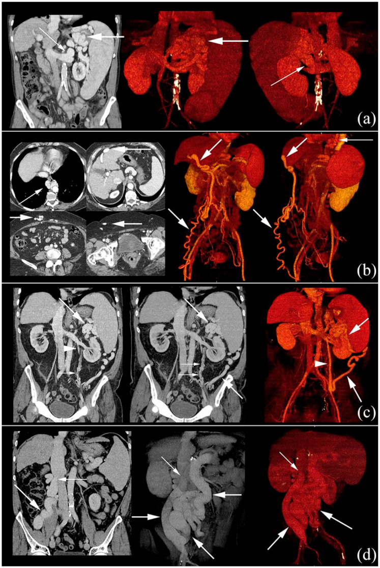
Figure 1.

Contrast-enhanced CTs of different SPSS.
(a) Gastrorenal shunt (thin arrows). Thick arrows: varices of coronary vein. Coronal image and two volume rendering images. (b) Paraumbilical shunt that drain through collaterals (thick arrows) to the right common femoral vein. Thin arrows: paraesophageal varices. Four axial images and two volume rendering images. (c) Splenorenal shunt (thick arrows) that communicates with left renal vein through left gonadal vein (arrowhead). Secondary peri-ureteral collaterals (thin arrows). Coronal image, maximum intensity projection coronal image and volume rendering image. (d) Mesocaval shunt (thick arrows), from SMV to IVC (thin arrows) through right gonadal vein. Coronal image and two volume rendering images.
CT: computed tomography; IVC, inferior vena cava; SMV, superior mesenteric vein; SPSS, spontaneous portosystemic shunt.
