
FIG. 1.
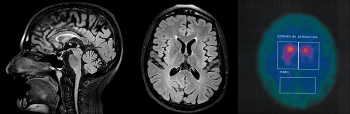
Magnetic resonance imaging and dopamine transporter scan of case 2. Sagittal (A) and transversal (B) fluid attenuated inversion recovery magnetic resonance imaging sequences show generalized brain atrophy with prominent parietal and cerebellar involvement as well as an “ear of the lynx sign” on the left (B). Dopamine transporter single‐photon emission computed tomography demonstrating reduced tracer uptake predominantly on the left.