Figure 1.
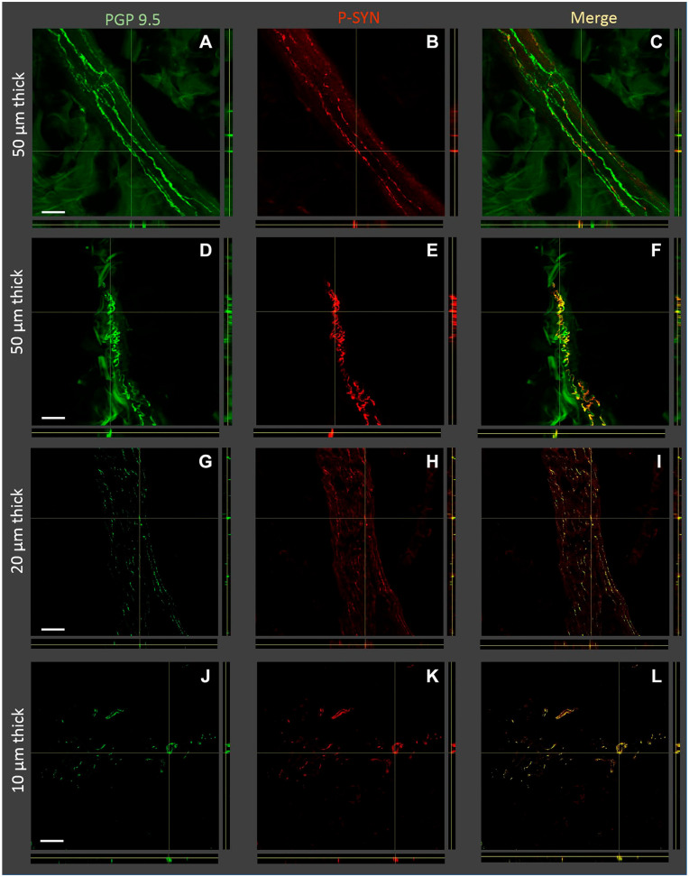
Orthogonal Z-stack images of tissue sections with nerve fibers containing phosphorylated alpha-synuclein (P-syn) in individuals with Parkinson’s disease. Four examples of tissue sections are shown: two examples of 50 µm thickness, one example of 20 µm thickness, and one example of 10 µm thickness (displayed on the left Y-axis). The immunostains used for each image are shown on the top of the figure and included protein gene product 9.5 (PGP 9.5), P-syn, and the merged images. Sections A–C and G–I show pilomotor nerve fibers, and sections D–F are a nerve bundle. Sections J–L contain both pilomotor nerve fibers and nerve bundles. Thicker tissue sections tend to allow for easier identification of overlapping PGP 9.5 and P-syn. Scale bar, 100 μm.

