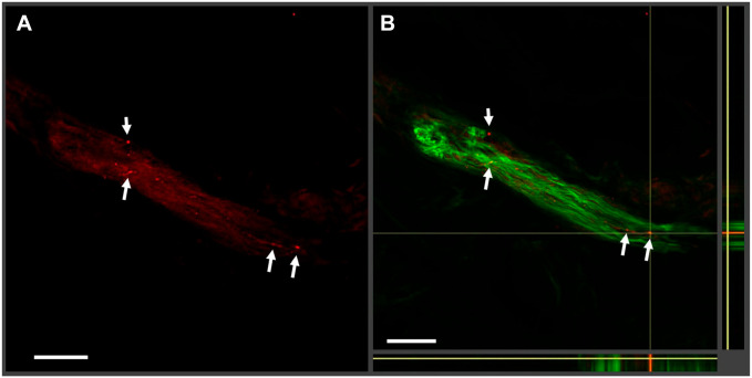
Figure 2.
Examples of phosphorylated alpha-synuclein (P-syn) artifacts. In this 50-µm-thick tissue section, a nerve bundle is stained with P-syn (seen in red in (A)) and protein gene product 9.5 (PGP 9.5 in green) with the merged orthogonal image shown in (B). The white arrows indicate regions of possible P-syn deposition in red. In the merged image, the regions do not colocalize with PGP 9.5. Without colocalization, it is easy to misinterpret artifacts for actual alpha-synuclein staining.

