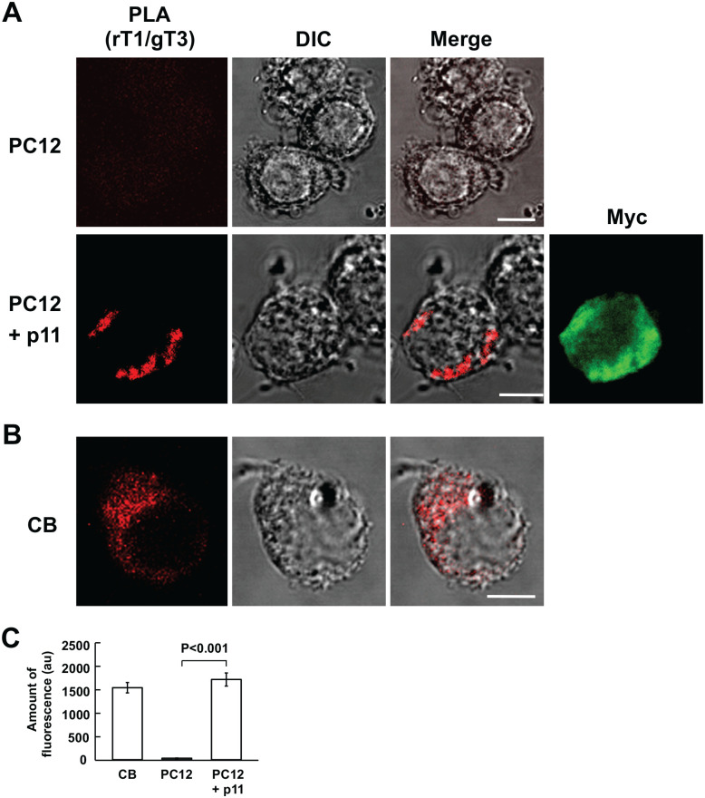
Figure 3.
Proximity ligation assay for heteromeric TASK1-TASK3 channel formation in PC12 cells and CB glomus cells. (A and B) Confocal images of PLA products in wild (PC12) and p11-expressing PC12 (PC12 + p11) cells and CB cells, respectively. First column represents fluorescence images of PLA products; second, DIC images; third, merge of fluorescence and DIC images; fourth, myc-like fluorescence image; bars are 5 µm. (C) Summary of fluorescence of PLA products in CB cells, and wild (PC12) and p11-expressing PC12 cells (PC12 + p11); data are mean ± SEM; Student’s t test. (CB: n=6 from two CBs; PC: n=6 from two culture dishes; PC12 + p11: n=6 from two culture dishes). PC12 cells were stimulated with NGF for 1 day (p11-expressing PC12 cells). Bars are 5 µm. Abbreviations: CB, carotid body; DIC, differential interference contrast; PC, pheochromocytoma; PLA, proximity ligation assay; NGF, nerve growth factor; TASK, TWIK-related acid-sensitive K+.

