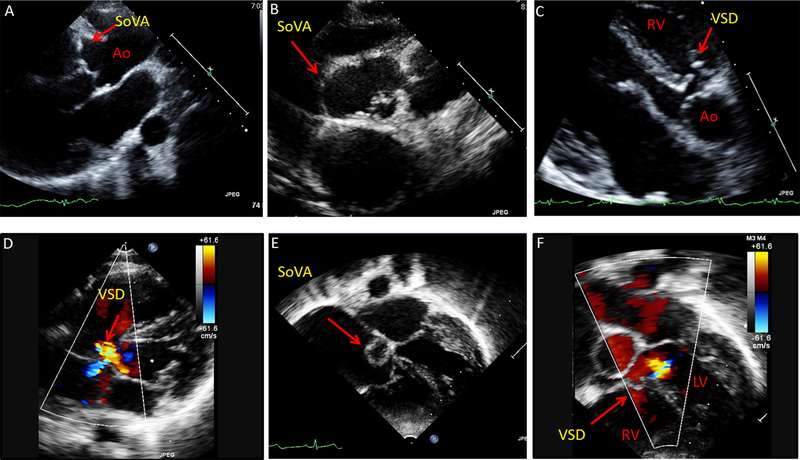
Figure 4: Sinus of Valsalva aneurysms (SoVA) with perimembranous ventricular septal defect in 2 young females.
(A-B) Echocardiography images from 21 year-old female with SoVA involving the right and non-coronary cusps of the aorta (parasternal long-axis and short-axis views). (C-D) Patient also has a perimembranous ventricular septal defect (VSD) with shunt flow, seen on parasternal and apical views. (E-F) Echocardiography images from an 11 year-old female depicting sinus of Valsalva aneurysm and a perimembranous VSD (apical 5-chamber view). Ao = aorta, LV = left ventricle, RV= right ventricle.

