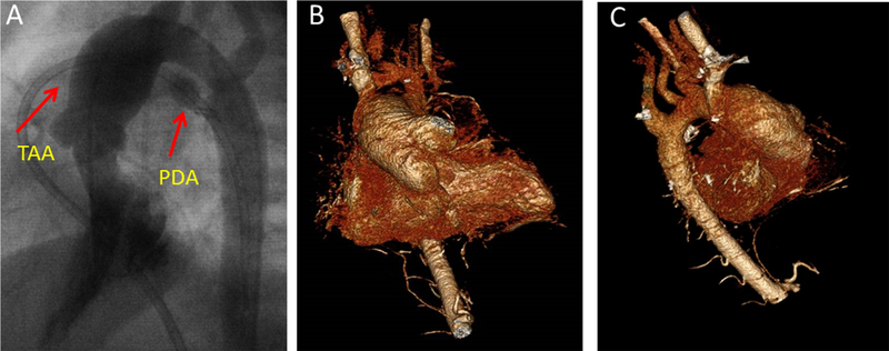
Figure 6: Female with progressive thoracic aortic dilatation requiring surgery.
(A) Dilated aorta and PDA seen on cardiac catheterization images during infancy. (B-C) 3D CT images of the patient’s aorta, obtained a decade later, demonstrate persistent dilatation of the aortic root and ascending aorta without involvement of the distal arch or descending aorta.

