Figure 3.
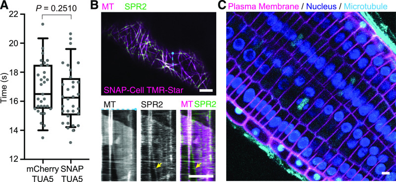
SNAP-Tagging Did Not Have a Significant Impact on the Duration of Mitotic Cell Divisions and Enabled in Vivo Multicolor Imaging in Arabidopsis Seedlings.
(A) Comparison of the duration from nuclear envelope breakdown to the phragmoplast initiation between mitotic cells in mCherry-TUA5 plants (n = 31) and SNAP-TUA5 plants incubated in 500 nM SNAP-Cell TMR-Star (n = 32). There was no significant difference (P = 0.2510 by Mann-Whitney U test). In the box plots, the boxes represent the range from the 25th to 75th percentiles, the horizontal lines represent the median value, and the whiskers span from the 5th to 95th percentiles.
(B) Cotyledon epidermal cells of Arabidopsis coexpressing SPR2-GFP (SPR2) and pUBQ10:SNAP-TUA5 (MT). Four-day-old seedlings were incubated in 0.5× MS containing 500 nM SNAP-Cell TMR-Star. (Top) Representative confocal image of MTs and SPR2 in a pavement cell. (Bottom) Kymographs generated from the top panel (at the blue dotted line) showing SPR2 tracking the minus end of microtubule (yellow arrow).
(C) Root tip of Arabidopsis coexpressing p35S:YFP-LTI6b, p35:H2B-RFP, and pUBQ10:SNAP-TUA5. Three-day-old seedlings were incubated in 0.5× MS containing 500 nM SNAP-Cell 647SiR for 30 min. Bars = 5 µm. Experiments were repeated independently two times ( [A] and [B]) and three times (C)) with comparable results.

